Table of Contents

What is Centriole?
All animal cells have two centrioles. They help the cell during cell division. They work during the process of mitosis and meiosis. It can be found in other low-lying plants such as Chlamydomonas, although not in many fungi, angiosperms (flowering plants), and pinophyta (conifers). They are usually located near the nucleus but are not visible when the cell does not separate.
Centrioles Structure
All centrioles are composed of 9 groups of microtubule triplets arranged in a cylindrical pattern. The detailed structure of centrioles can only be read with an electron microscope. These are linked together at right angles to each other.
Embryos of Drosophila melanogaster and C. elegans are different from this organization. The former made 9 pairs instead of microtubule triplets, while the premature embryos and C-sperm. elegans has 9 microtubule ones.
Edouard van Beneden and Theodor Boveri observed and detected centrioles for the first time in 1883 and 1888. The structure of multiplying centrioles was first introduced by Joseph G. Gall and Etienne de Harven in the 1950s.
Centriole helps to regulate the mitotic spindle and completes the cytokinesis process. However, centrioles were believed to be necessary for the formation of a mitotic spindle in the animal cell. Although, a few recent studies have suggested that a cell that does not have a centriole (laser-extracted) can function without it at the G1 interphase level and can be later constructed in the form of de novo.
The location of centrioles plays an important role in the three-dimensional organization of the cell as it also controls the location of the nucleus.
In flagellated and ciliated animals the location of such organelle is determined after the mother centrioles form the base.
Centrioles Function
The following is the important function of centrioles:
Despite their lack of DNA, the centrioles are able to form new centrioles.
They can be converted into basal bodies.
Basal bodies produce flagella and cilia.
They help to divide cells by building microtubule editing centers.
At two centrioles, the distal centriole forms a tail or axial filament.
Mobile Organization Standards
Cell Membrane:
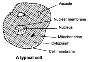
This membrane acts as a relatively inaccessible barrier or can also be called a barrier that allows very few particles in it while blocking most of the naturally occurring chemicals inside the cell.
Cell Walls:
Not all living things have cell membranes, especially animals and painters like animals. The cell walls contain viruses that include the chemical peptidoglycan. Cellulose, a non-digestible polysaccharide (in humans) is a common chemical within the wall of voltaic cells of a plant. Some plant cells similarly contain lignin and additional chemicals embedded within the cell walls of storage.
Nucleus:
In eukaryotic cells, only the nucleus can be found similar to the nucleic acids produced by RNA and DNA cells. RNA, also called Ribonucleic acid, is formed within the nucleus by means of DNA-based sequences as a prototype. RNA emerges from within the cytoplasm where it aids in protein synthesis. The nucleolus can be the part of the nucleus where ribosomes are formed.
Vacuoles and Vesicles:
Types of organelles that have a single membrane (also known as tonoplast) and are located inside the cell. In many creatures, vacuoles are the last resort. The vesicles are responsible for carrying substances from the inside of the cell to the outside.
Ribosomes:
Ribosomes are pigments that form proteins that do not have an outer membrane and are found in both eukaryotes and prokaryotes. Eukaryotic ribosomes are larger than prokaryotic cells. The ribosome contains a small and large units. Biochemically, the ribosome contains rRNA (ribosomal RNA) and a few 50 structural proteins.
Endoplasmic Reticulum:
The Endoplasmic reticulum is a network of connective tissue that assists in the transport and binding of proteins. Rough ER (Rough endoplasmic reticulum) got its name because of its rough outer surface due to the presence of several ribosomes found on the outer endoplasmic reticulum. The Smooth Endoplasmic Reticulum has no ribosomes.
Golgi Apparatus:
Golgi Apparatus or structures were first discovered in the 1890s by Italian Italian biologist Camillo Golgi. They are compressed stacks of membrane-bound sacs that help to regenerate vesicles produced by the endoplasmic reticulum.
Lysosomes:
Larger vesicles are composed of Golgi. They incorporate hydrolytic enzymes that will release cells. Lysosome content is inherited to use within extracellular fractures.
Mitochondria:
Found in eukaryotic cells that contain their DNA. Their help is due to the site of energy release and ATP formation (in the form of chemiosmosis). The mitochondrion is labeled because of its power cell. Mitochondria have two membranous valleys. The formation of ATP (Adenosine triphosphate) occurs in the folding cell wall of mitochondria, also known as cristae. The inner membrane surrounds the mitochondrion matrix containing mitochondrial DNA and ribosomes.
Mitochondria and Endosymbiosis:
The concept of endosymbiosis was proposed by Lynn Margulis in the 1980s. This concept describes the chloroplasts and mitochondria of prokaryotes.
Plastids are organelle present in plants and photosynthetic eukaryotes and bound to the membrane. Leucoplasts are also considered starchy amyloplasts and sometimes fats or proteins. Chromoplasts keep pigs associated with the attractive color of flowers or fruits.
Also read: Cilia
FAQs
1. What is the function of centrioles?
Ans: The centriole is the organelle in the animal cell. These are tube-like structures and are made of tubulin protein and are an integral part of centrosomes. These are mainly found in wet animal cells, fungi, and algae but not in plants. This enemy is a major component of eukaryotic cells. These are located very close to the nucleus of the cell.
Functions of centrioles
The functions of centrioles are:
The main function of the centriole is to assist in cell division in animal cells.
Centrioles also contribute to the formation of spinning strands that separate chromosomes during cell division (mitosis).
The second function of the centrioles we will focus on is celiogenesis. Celiogenesis is the formation of cilia and flagella on the surface of cells. Cilia and flagella aid in cell mobility.
2. Do plant cells have centrioles?
Ans: Centrioles can be found at:
- Animal cells
- Low plants
- Base cilia and flagella (as basic bodies)
- Centrioles are commonly found in eukaryotic cells, not in high-grade plants. In these plants, cells do not use centrioles during cell division. The lower flagella plant has a centriole at the base of the flagella and supports cell division and cell movement.


