
Table of Contents
Introduction
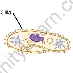
The cilia is a tiny protuberance that extends beyond the eukaryotic cells from a very wide cell body.
Cilia is divided into two types, namely: cilia in motile and non-motile. The primary cilia are non-motile cilia that act as nerve organelles. The single main stagnant cilium acts as a cell membrane for certain types of mammal cells. The exceptions include odorous neurons, which have multiple cilia, and temporary embryonic node cells, with a single motile cilia called nodal cilia, which are important in the development of left-right body asymmetry.
Motile cilia and flagella (commonly called undulipodia) were similar in structure to eukaryotes, however, segregation is usually based on function or length. The primary cilia (cilia immotile) transmit signals from space or other cells.
Cilia Genres
Basic Cilia:
Non-motile cilia can be present in almost every cell in the animal kingdom, and blood cells have become remarkable. Compared with cells containing motile cilia, some cells have only one, probably other than the olfactory nerve neurons, which have about ten cells and contain fragrant receptors.
When the main cilium was identified in 1898, these were largely ignored during the next century, and it was thought that the investigative organelle had no significant role. Its physiological functions in signal transfer, chemosensation modification, and control of cell growth have only recently been discovered, highlighting its importance in cell function. The discovery of its presence in a variety of diseases associated with dysgenesis or cilia deficiency, including congenital heart disease, polycystic kidney disease, and retinal degeneration, known as ciliopathies, also demonstrated its importance in human biology. Several human organs now have a primary cilium that plays a key role in their function.
Motile Cilia: Motile cilia cells are also found in large eukaryotes, such as mammals. Motile cilia are found in large numbers on the surface of cells and are thus struck by symmetrical waves.
Motile cilia are found in the respiratory epithelium which focuses on the respiratory tract in humans, where they help with mucociliary excretion, which includes the clearing of mucous membranes and mucus from the lungs. The respiratory epithelium contains approximately 200 motile cilia per cell.
The ovum is transferred from the uterus to the uterus through the cilia of the oviduct in female mammals.
Epithelial cells including their choroid plexus epithelial cells have motile cilia. These are found in large numbers in each cell and move slowly, placing them between the motile and the primary cilia. Within the hair cells, there was a 9 + 2 cilia that did not move over the 9 + 0 cilia that were moving.
Maintaining high levels of periciliary fluid to wash the cilia is essential for the motile cilia to function properly. The sodium channels of the ENaC epithelial, which are distributed throughout the entire length of the cilia, appear to act as nerves that control the amount of fluid that covers the cilia.


Nodal Cilia
The third form of cilium is nodal cilium, which is motile 9 + 0 cilium. Nodal cilia usually occur during early embryonic development. It has no intermediate material, however, it has dynein arms that allow it to move or rotate in a circular motion, similar to the old cilium. The circular motion of the nodal cilium causes the movement of extraembryonic fluid throughout the node, directed to the left. The directional flow feels the primary cilia across the nodal cilia, which stimulates the nodal signature and establishes the left to the right side.
What is Cilia Anatomy?
The axoneme appears to be the microtubule-based cytoskeleton found between the cilia and the flagella. The axoneme of that main cilium usually consists of a ring of nine outer microtubulees (called a 9 + 0 axoneme), while the moving axoneme of the cilium consists of two medium-sized singlets as opposed to a double double double axis (called axoneme 9 + 2).
The axoneme acts as a scaffold for the axonemal anterior and posterior dynein arms, which transmit motile cilia cells, and tracks molecular motor proteins such as Kinesin II, which transport proteins throughout the length of the cilium by intraflagellar transport (IFT). IFT appears to be a bi-directional, retrograde device. To return to the cell body, IFT uses the cytoskeletal dynein motor 2. Cilium is trapped by a membrane similar to that of plasma.
Ciliary Rootlet
A cytoskeleton-like called ciliary rootlet structure that extends from the basal end of the cilium body. Cross striae are propagated at intervals of about 55-70 nm in rootlets usually 80-100 nm wide. Rootletin is a key component of rootlet.


Transition Zone
The closest-most area of the cilium has a transition zone responsible for controlling which proteins must combine and leave the cilium to form a different structure. Y-shaped structures bind to the ciliary membrane in the lower axoneme throughout the evolutionary region. A feature such as a transition area filter can be used to monitor selected entry into the cilia.
Also read: Flagella
Cilia Versus Flagella
Motile cilia and flagella, despite their various names, have similar structures and serve the same purpose: movement. The appendage movement can be compared to an earthquake. Wave is defined by amplitude, frequency (ciliary beat frequency (CBF)), and wavelength, and appears to originate from the base of the cilium. The cilia beat movement is caused by a double external slide by the arm structures of the dynein, and originates in the axoneme rather than the basal body. The main difference between the two systems is that the flagella drive the eukaryotic organisms like humans, while the cilia transmit substances into space.
Cilia Use
Axoneme’s dynein forms bridges between adjacent double microtubules. Once ATP activates the dynein motor vehicle, it attempts to cut a microtubule doublet near it. If it were not for the presence of Nexin between the double microtubules, it would cause nearby adjusts to slide over one another. As a result, the energy produced by dynein is converted into a bending motion.


Sensitive Outer Environment
In eukaryotes, a few primary cilia near epithelial cells act as cellular horns, sensing the extracellular outer area for thermosensation, chemosensation, and mechanosensation. Such cilia, therefore, link various signals, such as dissolving elements throughout the outer cell, the secretory components in which soluble protein is produced to provide a lower effect of fluid flow and to mediate the flow of water when the cilia become motile.
Most epithelial cells melt, and these usually form a tube or tube through which the cilia extend as just as a layer of bright cells. Cilia play an important role in sensory and signal storage in the cellular environment, which may explain why ciliary dysfunction produces a variety of human diseases.
