Banner 0
Banner 1
Banner 2
Banner 3
Banner 4
Banner 5
Banner 6
Banner 7
Banner 8
Banner 9

Q.

What does an endoplasmic reticulum look like?

see full answer

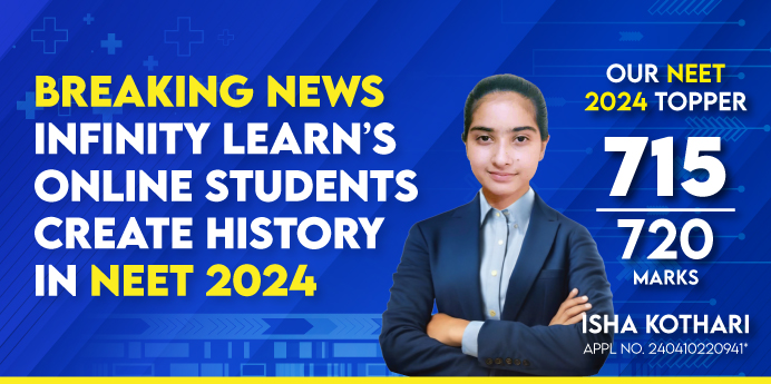
High-Paying Jobs That Even AI Can’t Replace — Through JEE/NEET

🎯 Hear from the experts why preparing for JEE/NEET today sets you up for future-proof, high-income careers tomorrow.
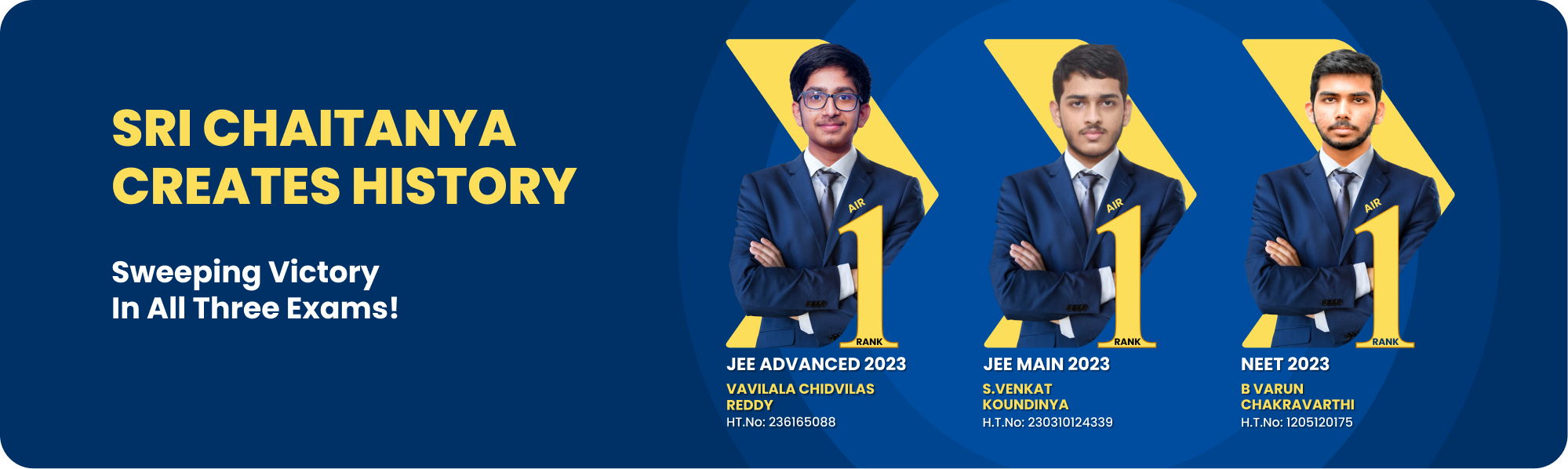
An Intiative by Sri Chaitanya

(Unlock A.I Detailed Solution for FREE)

Best Courses for You

JEE

JEE

NEET

NEET

Foundation JEE

Foundation JEE

Foundation NEET

Foundation NEET

CBSE

CBSE

Detailed Solution

The endoplasmic reticulum (ER), when visualized under an electron microscope, appears as a massive, intricate network of membranes that spreads throughout the cytoplasm of eukaryotic cells. It resembles a highway system within the cell, interconnecting various regions and facilitating transport and communication between the nucleus and other organelles. This membrane-bound structure is continuous with the outer nuclear membrane, giving it a strategic position to manage and direct cellular traffic.

Appearance of the Rough and Smooth ER

The ER can be broadly classified into two types based on appearance:

  • Rough Endoplasmic Reticulum (RER):
    • The RER looks rough or bumpy due to the presence of ribosomes attached to its cytoplasmic side.
    • Under high-resolution microscopes, it resembles flattened, sheet-like cisternae (or sacs), often stacked and arranged in parallel lines.
    • This rough appearance signifies its role in protein synthesis and is generally located near the nucleus.
  • Smooth Endoplasmic Reticulum (SER):
    • The SER has a smooth, tubular appearance since it lacks ribosomes.
    • It consists of branching, interlinked tubules, forming a network that looks like intertwined pipes or threads.
    • It is primarily involved in lipid metabolism, detoxification, and calcium storage.

Colors and Diagrammatic Representation

In textbook diagrams, the ER is usually depicted in bright colors:

  • Rough ER is shown with small dots on its surface representing ribosomes.
  • Smooth ER is illustrated as plain, looping tubules without any surface attachments.

Though cells do not have inherent colors, these schematic representations help students understand the structural differences between the two types of ER.

Advanced Microscopy Techniques

Scientists use:

  • Electron Microscopes to reveal the ultrastructure of ER in fine detail.
  • Fluorescence Microscopy with staining dyes to visualize ER in live cells, where it appears as a glowing network dynamically moving within the cytoplasm.

Dynamic Nature of ER

A fascinating aspect of ER’s appearance is its dynamic behavior:

  • It is not static but continuously reshapes itself, expanding and retracting based on the cell’s metabolic activity.
  • During cell division, the ER network is restructured to ensure proper distribution to daughter cells.

Summary

The endoplasmic reticulum looks like a highly branched, continuous network of membranes, with RER appearing rough due to ribosomes and SER appearing smooth with tubular structures. It functions as the cell’s internal transport and production hub, contributing directly to the synthesis and distribution of essential molecules.

Watch 3-min video & get full concept clarity

Ready to Test Your Skills?

Check your Performance Today with our Free Mock Test used by Toppers!

Take Free Test

score_test_img

Get Expert Academic Guidance – Connect with a Counselor Today!

whats app icon
What does an endoplasmic reticulum look like?