- Tissues in Action Class 9: Chapter Overview
- Download Tissues in Action Class 9 Notes PDF
- Why are Plant and Animal Tissues Different?
- What is a Tissue?
- Plant Tissues
- Permanent Tissues
- Types of Permanent Tissues
- Xylem vs Phloem
- Plant Tissue Systems
- Animal Tissues
- Types of Animal Tissues
- Muscular Tissue
- Skeletal Muscle
- Smooth Muscle
- Cardiac Muscle
- The Musculoskeletal System
Tissues in Action Class 9 Notes
By Karan singh ildefaultfield
|
Updated on 8 Sep 2026, 16:10 IST
Tissues in Action Class 9 Notes provide a detailed explanation of the important concepts covered in Class 9 Science Chapter 3. These notes help students understand plant tissues, animal tissues, their types and functions, the musculoskeletal system, joints, totipotency, and crown gall disease in a simple and easy-to-revise format.
The notes include important definitions, explanations, comparison tables, diagrams, and key points to help students understand the chapter and prepare for their examinations. Students can read the topic-wise explanations below and use the downloadable PDF for quick revision and offline study.
Tissues in Action Class 9: Chapter Overview
In this chapter, students learn about how cells are organised into tissues and how different tissues perform specialised functions. The chapter covers both plant and animal tissues, along with their structures and functions.
The major topics include:
- What is a tissue?
- Plant tissues
- Meristematic tissues
- Permanent tissues
- Parenchyma, collenchyma and sclerenchyma
- Xylem and phloem
- Plant tissue systems
- Epidermis and stomata
- Animal tissues
- Epithelial tissue
- Connective tissue
- Muscular tissue
- Nervous tissue
- Musculoskeletal system
- Types of joints
- Skeletal system
Download Tissues in Action Class 9 Notes PDF
Why are Plant and Animal Tissues Different?
Plants and animals are both made up of cells, but their tissues differ because they have different lifestyles and perform different functions. Plants remain fixed in one place and therefore require strong supporting tissues to keep them upright. Animals, in contrast, move from place to place, so they need flexible tissues that allow movement.
Their mode of nutrition is also different. Animals obtain food from various sources and have specialised tissues that help in digestion and absorption. Plants, however, make their own food. Their tissues help capture and use sunlight to produce food through photosynthesis.
Loading PDF...
| Basis | Plant Tissues | Animal Tissues |
| Cell wall | Present | Absent |
| Growth | Growth is limited to specific regions called meristems | Growth occurs in many parts of the body |
| Movement | Plants remain fixed in one place, so their tissues mainly provide support | Animals move actively, so many tissues are specialized for movement |
| Living or dead cells | Many supporting tissues contain dead cells | Most tissues are made of living cells |
| Energy requirement | Generally require less energy | Generally require more energy |
| Types of tissues | Meristematic tissues and Permanent tissues — Simple: Parenchyma, Collenchyma, Sclerenchyma; Complex: Xylem, Phloem | Epithelial, Connective, Muscular, and Nervous tissues |
| Main functions | Support, transport, storage, and photosynthesis | Protection, movement, coordination, and transport |
| Flexibility | Generally less flexible | Generally more flexible |
What is a Tissue?
A tissue is a group of similar cells that work together to perform a particular function. In multicellular organisms, cells become specialised for different activities. Groups of specialised cells form tissues, tissues combine to form organs, and organs work together as organ systems.
Organisation in multicellular organisms
Cell
↓
Tissue
↓
Organ
↓
Organ System
↓
Organism
For example:
- Muscle cells form muscular tissue.
- Muscular tissue helps in movement.
- Nervous tissue helps transmit information.
- Xylem transports water and minerals in plants.
Thus, tissues allow division of labour, making the functioning of complex organisms more efficient.

Plant Tissues
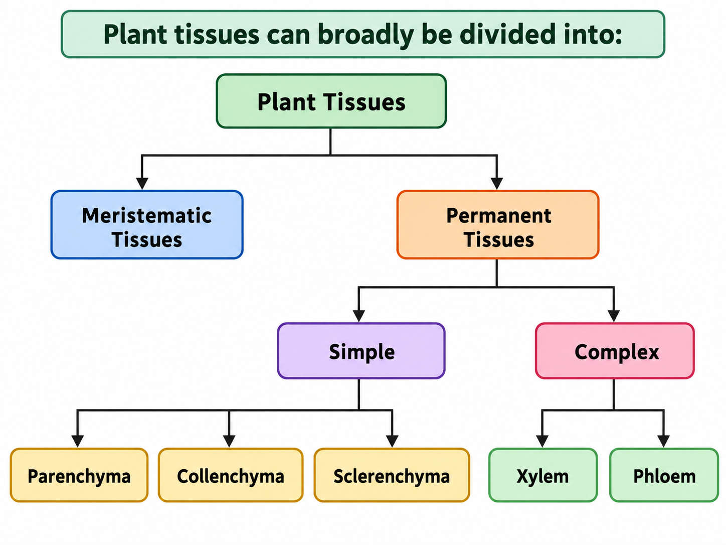
Plants have specialised tissues that help them grow, provide support, transport substances, store food, and perform photosynthesis. Plant tissues can broadly be divided into:
Best Courses for You

JEE

NEET

Foundation JEE

Foundation NEET

CBSE
Meristematic Tissue
Meristematic tissue is made up of cells that actively divide and help in the growth of plants. Plants continue to grow because new cells are continuously produced by meristematic tissues.
Main characteristics
Meristematic cells generally have:
- Small size
- Thin cell walls
- Dense cytoplasm
- Large and prominent nucleus
- Many cell organelles
- Little or no intercellular space
- Generally no large vacuoles
These features enable the cells to divide repeatedly.
Types of Meristematic Tissue
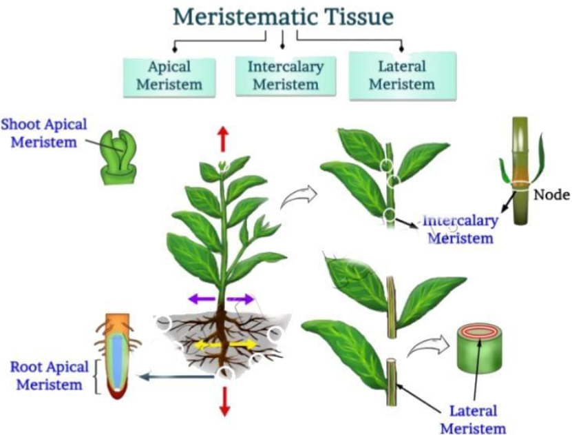
There are three major types:
- Apical meristem
- Lateral meristem
- Intercalary meristem
Apical meristem
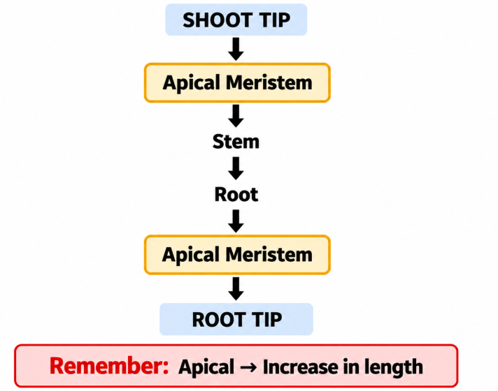
Apical meristem is a type of meristematic tissue made up of young cells that divide continuously. It is mainly responsible for the increase in length of the plant. Apical meristem is present at the growing tips of roots and shoots. In roots, it is found just behind the root cap. In shoots, it is present at the tip of the stem and in growing buds. It brings about primary growth, which increases the length of roots and shoots. In roots, it helps the root grow deeper into the soil. In shoots, it helps the stem grow upward. It also produces new cells that later differentiate into tissues and organs such as leaves, stems, and branches.
Diagram
Lateral Meristem
Lateral meristem is a type of meristematic tissue that helps a plant increase in thickness or girth. Its cells divide continuously and produce new tissues toward the inside and outside of the stem and root. Lateral meristem is found along the sides of stems and roots. The main lateral meristems are vascular cambium and cork cambium.
It is responsible for secondary growth, which increases the diameter of stems and roots. The vascular cambium forms new xylem and phloem, while the cork cambium produces protective outer tissues. It also helps woody plants develop stronger and thicker stems.
Remember: Lateral meristem → Increase in thickness or girth.
Annual Growth Rings
The activity of lateral meristem can result in visible growth rings in tree trunks.
These rings can provide information about:
- The age of a tree
- Periods of favourable growth
- Periods of unfavourable growth
Intercalary Meristem
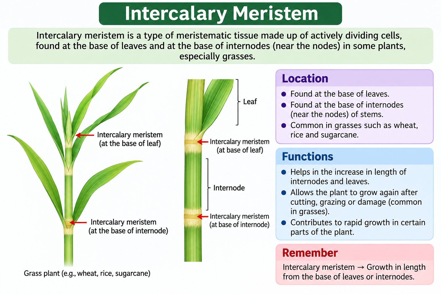
Intercalary meristem is a type of meristematic tissue made up of actively dividing cells. It helps certain parts of a plant continue to grow even after the tip has stopped growing. Intercalary meristem is usually found at the base of leaves and at the base of internodes or near the nodes of stems. It is especially common in grasses, wheat, rice, and sugarcane. Its main function is to increase the length of internodes and leaves. It also helps grasses and similar plants regrow quickly after cutting, grazing, or damage because the growing tissue is located near the base rather than only at the tip.
Remember: Intercalary meristem → growth in length from the base of leaves or internodes.
Permanent Tissues
When a stem, root, or leaf is observed under a microscope, the cells are seen to differ in shape, size, and function. Groups of these specialised cells form permanent tissues. Permanent tissues are made up of mature cells that have lost the ability to divide and have become specialised to perform particular functions.
Permanent tissues are of two main types:
- Simple permanent tissues – made up of only one type of cell
- Complex permanent tissues – made up of more than one type of cell
Why are permanent tissues formed?
Permanent tissues are formed when meristematic cells mature, stop dividing, and become specialised for particular functions. This process is called differentiation. Permanent tissues are needed because different parts of a plant perform different jobs, such as storage, photosynthesis, support, protection, and transport of water and food. For example, some permanent cells become specialised to store food, while others form tissues such as xylem and phloem for transport.
In short: Meristematic cells → differentiation → permanent tissues → specific functions
Meristematic Tissue vs Permanent Tissue
| Basis | Meristematic Tissue | Permanent Tissue |
| Definition | Tissue made of actively dividing cells | Tissue made of mature cells that have lost the ability to divide |
| Cell division | Cells divide continuously | Cells generally do not divide |
| Cell size | Cells are usually small and compact | Cells are generally larger and fully developed |
| Cell wall | Thin primary cell wall | Cell wall may be thin or thick depending on the tissue |
| Cytoplasm | Dense cytoplasm | Less dense cytoplasm |
| Nucleus | Large and prominent | Usually smaller compared with meristematic cells |
| Vacuoles | Vacuoles are absent or very small | Large vacuoles are usually present |
| Intercellular spaces | Usually absent | May be present |
| Main function | Helps in plant growth by producing new cells | Performs specific functions such as support, storage, photosynthesis, protection, and transport |
| Location | Found in growing regions such as root tips, shoot tips, and cambium | Found in mature parts of roots, stems, and leaves |
| Examples | Apical, lateral, and intercalary meristems | Parenchyma, collenchyma, sclerenchyma, xylem, and phloem |
Types of Permanent Tissues
There are two main types of permanent tissues in plants:
(i) Protective Tissue - Epidermis
The epidermis forms the outermost protective covering of the plant. It covers the leaves, stems, and roots and is generally made up of a single layer of closely packed cells. In many plant parts, the epidermis is covered by a waxy layer called the cuticle, which helps protect the plant.
Functions of Epidermis:
- Protects the plant from mechanical damage and harmful microorganisms.
- Helps reduce water loss through transpiration.
- In roots, some epidermal cells develop into root hairs, which increase the surface area for absorbing water and minerals.
(ii) Supporting Tissue - Simple Permanent Tissues
Simple permanent tissues are made up of similar types of cells and mainly help provide strength, support, and flexibility to different parts of the plant.
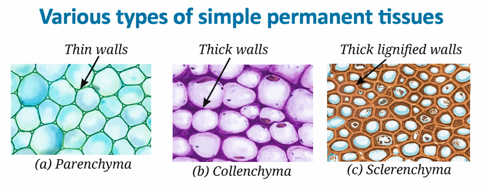
They are of three types:
- Parenchyma
- Collenchyma
- Sclerenchyma
a. Parenchyma
Parenchyma is made up of living cells with thin cell walls. The cells are loosely arranged, leaving spaces between them. Its primary function is to store food, while in green parts of plants, it also helps carry out photosynthesis. In aquatic plants, specialised parenchyma develops large air spaces that help the plants float on water.
b. Collenchyma
Collenchyma consists of living cells whose cell walls are unevenly thickened at the corners due to the deposition of cellulose and pectin. This tissue provides mechanical support and flexibility, allowing plant parts such as young stems and tendrils to bend without breaking.
c. Sclerenchyma
Sclerenchyma is made up of cells with thick, strong walls due to the deposition of lignin. Most sclerenchyma cells are dead when mature. This tissue provides strength, rigidity, and support to the plant and is commonly found in stems, leaf veins, and the hard coverings of seeds and nuts, such as coconut husk and walnut shells.
(iii) Conducting tissues - Complex permanent tissues
Conduction tissues in plants are specialised tissues that help transport water, minerals, and food throughout the plant body. They are known as complex permanent tissues because they are made up of different types of cells that work together to perform transport functions.
Types of Conducting Tissues
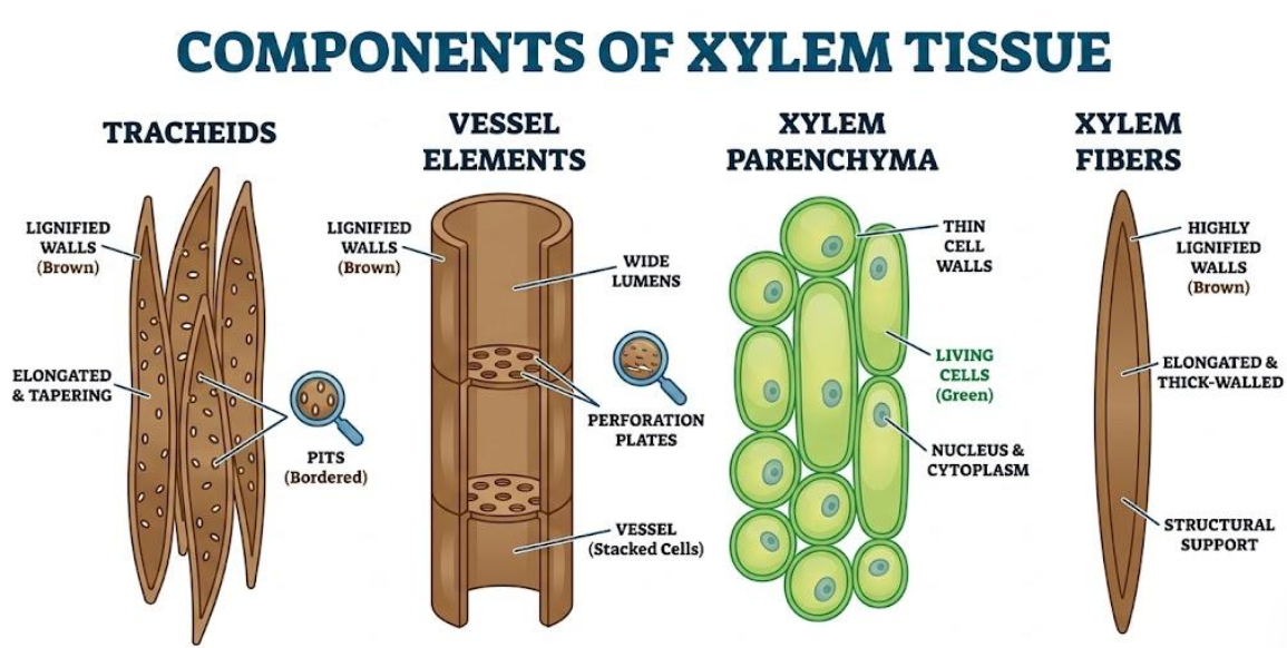
1. Xylem
Xylem is a complex permanent tissue that transports water and minerals from the roots to the leaves and other parts of the plant. It also helps provide mechanical strength and support to the plant.
- Tracheids and vessels are long, tubular structures with thick walls that help conduct water.
- Most xylem cells are dead at maturity, except xylem parenchyma, which is living.
- Xylem mainly helps in the upward transport of water and minerals through the plant.
Components of Xylem
- Tracheids
- Vessels
- Xylem parenchyma
- Xylem fibres
2. Phloem
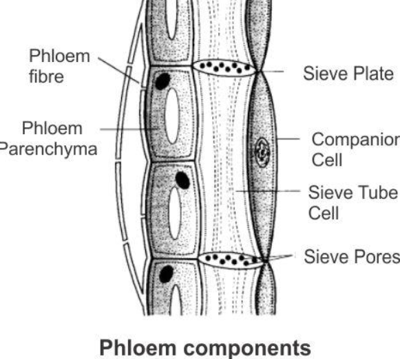
Phloem is a complex permanent tissue that transports food, mainly sugars, produced in the leaves to other parts of the plant.
- Phloem is composed mainly of living cells.
- Sieve tubes are long, tubular cells arranged end-to-end with perforated walls that facilitate food transport.
- Companion cells help regulate the functioning of the sieve tubes.
- Phloem parenchyma helps in the storage of food and other substances.
Components of Phloem
- Sieve tubes
- Companion cells
- Phloem parenchyma
- Phloem fibres
Xylem vs Phloem
| Xylem | Phloem |
| Transports water and minerals | Transports food |
| Conducting tissue of water/minerals | Conducting tissue of food |
| Contains tracheids and vessels | Contains sieve tubes and companion cells |
| Also provides support | Phloem fibres provide support |
Plant Tissue Systems
Plant tissue systems are groups of tissues that work together to perform important functions in a plant. They are mainly divided into three tissue systems: dermal, ground, and vascular tissue systems.
1. Dermal Tissue System
The dermal tissue system forms the outer protective covering of the plant. It is found on the outer surface of roots, stems, and leaves. It protects the plant from injury and water loss, helps prevent the entry of harmful microorganisms, and in roots it helps absorb water and minerals through root hairs. Examples: Epidermis, root hairs, and cork.
2. Ground Tissue System
The ground tissue system makes up most of the internal part of the plant body between the dermal and vascular tissues. It performs photosynthesis, food storage, and support. Main tissues:
- Parenchyma – storage and photosynthesis
- Collenchyma – flexible support
- Sclerenchyma – strong mechanical support
3. Vascular Tissue System
The vascular tissue system is responsible for the transport of materials throughout the plant. It carries water, minerals, and food from one part of the plant to another. It consists mainly of:
- Xylem – transports water and minerals from roots to other parts of the plant.
- Phloem – transports prepared food from leaves to the rest of the plant.
Animal Tissues
Like plant cells, animal cells also work together in groups and become specialised to carry out specific functions. A group of similar cells performing a particular function forms an animal tissue. Animal tissues help the body perform many important activities such as movement, breathing, sensation, protection, and coordination. You can observe the work of animal tissues through simple actions such as:
- Blinking your eyes
- Clenching and opening your fist
- Taking a deep breath
- Touching something warm or cold
Types of Animal Tissues
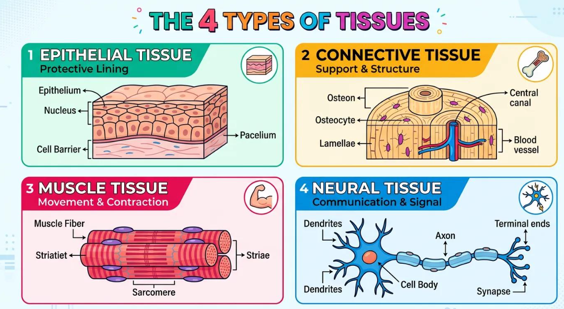
Epithelial Tissue
Epithelial tissue is the protective tissue that covers the outer surface of the body and lines the internal organs, cavities, blood vessels, and ducts. It also forms many glands.
Structure of Epithelial Tissue
Epithelial tissue is made up of closely packed cells with very little or no space between them. The cells are arranged in one or more layers, depending on their function. The cells rest on a thin supporting layer called the basement membrane, which separates the epithelium from the tissues below it.
Epithelial tissue does not contain blood vessels of its own. It receives nutrients by diffusion from the underlying tissues. Depending on the shape of the cells and the number of layers, epithelial tissue occurs in different forms such as squamous, cuboidal, columnar, ciliated, and glandular epithelium.
Functions of Epithelial Tissue
The main functions of epithelial tissue are:
- Protection: It protects the body and internal organs from injury, germs, and harmful substances.
- Absorption: In organs such as the small intestine, it helps absorb digested nutrients.
- Secretion: Glandular epithelium produces substances such as sweat, saliva, mucus, and enzymes.
- Excretion: It helps remove waste products, for example through the kidneys and sweat glands.
- Diffusion and exchange: Thin epithelial layers allow gases and other substances to pass through easily, such as in the lungs.
- Movement of substances: Ciliated epithelial cells help move mucus and particles along the respiratory tract.
- Sensation: Some epithelial cells contain sensory receptors that help detect touch, taste, and other stimuli.
Epithelium for Exchange
Epithelium for exchange is a very thin type of epithelial tissue that allows substances to pass quickly from one side to the other. It is mainly formed by simple squamous epithelium, which consists of a single layer of flat, thin cells.
Because the cells are only one layer thick, the distance for diffusion is very small. This makes the tissue well suited for the exchange of gases, nutrients, and waste materials. It is found in places where rapid exchange is needed, such as the air sacs (alveoli) of the lungs and the walls of blood capillaries.
In the lungs, it allows oxygen to diffuse from the air into the blood and carbon dioxide to diffuse from the blood into the air. In capillaries, it helps in the exchange of nutrients, gases, and waste products between the blood and body tissues.
Epithelium for Protection
Protective epithelium is epithelial tissue that forms a strong covering over body surfaces and lines organs that are exposed to friction, injury, germs, and harmful substances. It is usually made up of several layers of closely packed cells, so it is also called stratified epithelium. The outer cells may wear away due to friction, but new cells from the deeper layers continuously replace them.
Protective epithelium is found in areas that experience frequent wear and tear, such as the skin, mouth, oesophagus, and other body passages. It protects underlying tissues from mechanical injury and friction, prevents the entry of microorganisms and harmful substances, and in the skin it also helps reduce water loss. In the skin, the outer epithelial cells contain keratin, which makes the surface tough and water-resistant. This type is called keratinised stratified squamous epithelium.
Epithelium for Secretion
Secretory epithelium is a type of epithelial tissue specialised to produce and release substances needed by the body. It is commonly called glandular epithelium. The cells are closely packed and are modified for secretion. They may occur as single secretory cells or form groups of cells called glands.
It is found in glands such as the salivary glands, sweat glands, gastric glands, intestinal glands, and endocrine glands. It secretes substances such as mucus, saliva, sweat, digestive enzymes, and hormones. These secretions help in lubrication, digestion, temperature regulation, and control of body activities.
Sensory Epithelium
Sensory epithelium is a specialised type of epithelial tissue that contains sensory receptor cells. These cells detect changes or stimuli from the environment and help the body respond to them. Sensory epithelial cells are closely packed and are associated with nerve endings. They are modified to receive stimuli such as light, sound, smell, taste, and touch.
Sensory epithelium is found in sense organs, for example in the nose, tongue, inner ear, and parts of the eye. Its main function is to detect stimuli and transmit information to the nervous system. This helps the body recognise sensations such as smell, taste, sound, and other changes in the surroundings.
Epithelium for Absorption
Absorptive epithelium is a type of epithelial tissue specialised to absorb useful substances from one part of the body into the blood or surrounding tissues. The cells are usually thin and closely packed. In some organs, such as the small intestine, the cells have tiny finger-like projections called microvilli. These increase the surface area and help absorb substances more efficiently.
Absorptive epithelium is mainly found in the small intestine and in parts of the kidney tubules. It helps absorb digested nutrients, water, salts, and other useful substances. In the small intestine, it absorbs nutrients from digested food into the blood.
Connective Tissue
Connective tissue is a type of animal tissue that connects, supports, binds, protects, and transports materials between different parts of the body. Unlike epithelial tissue, the cells in connective tissue are usually widely spaced and are surrounded by a large amount of intercellular material called matrix. The nature of this matrix may be fluid, soft, flexible, or hard, depending on the type of connective tissue.
Connective tissue consists of cells, fibres, and matrix. The fibres provide strength and elasticity, while the matrix supports and holds the cells together. It helps to join tissues and organs, provide structural support, protect internal organs, store fat, transport substances, and repair damaged tissues.
Examples include:
- Blood
- Bone
- Cartilage
- Tendon
- Ligament
Blood
Blood is a fluid connective tissue.
It consists of:
- Plasma
- Red blood cells
- White blood cells
- Platelets
Functions
- RBCs: Transport oxygen with the help of haemoglobin.
- WBCs: Help defend the body against infection.
- Platelets: Help in blood clotting.
- Plasma: Provides the fluid medium in which blood cells and dissolved substances are transported.
Bone
Bone is a hard connective tissue.
Functions
- Provides support
- Gives strength
- Protects internal organs
- Forms part of the skeletal framework
Its matrix is hard and contains mineral substances including calcium and phosphorus compounds.
Cartilage
Cartilage is a connective tissue with a relatively soft and flexible matrix.
Functions
- Provides flexibility
- Cushions joints
- Absorbs shock
Examples
- Nose
- Ear
- Joints
Tendon
A tendon connects muscle to bone. When a muscle contracts, the tendon transmits the force to the bone and helps produce movement.
Ligament
A ligament connects bone to bone.
Functions
- Provides stability to joints
- Limits excessive movement
- Helps prevent dislocation
Tendon vs Ligament
| Tendon | Ligament |
| Connects muscle to bone | Connects bone to bone |
| Transmits force from muscle | Stabilises joints |
| Helps produce movement | Limits excessive movement |
Muscular Tissue
Muscular tissue is responsible for producing movement through contraction and relaxation.
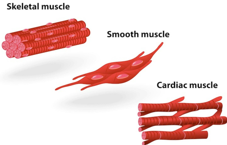
There are three types:
- Skeletal muscle
- Smooth muscle
- Cardiac muscle
Skeletal Muscle
Skeletal muscle is a type of muscle tissue mainly attached to the bones of the skeleton. It is responsible for most voluntary movements of the body, such as walking, running, writing, lifting objects, and changing facial expressions. Skeletal muscle works by contracting and relaxing. When it contracts, it becomes shorter and pulls on the bone to which it is attached, producing movement at a joint.
Structure of Skeletal Muscle
Skeletal muscle is made up of long, cylindrical cells called muscle fibres. These fibres are arranged parallel to one another and are grouped together to form muscles. Each skeletal muscle fibre has several important features:
- It is long and cylindrical.
- It is unbranched.
- It contains many nuclei, usually located near the outer edge of the cell.
- It shows alternating light and dark bands under a microscope. These bands are called striations, so skeletal muscle is also called striated muscle.
- Each muscle fibre contains many smaller structures called myofibrils.
Myofibrils are made mainly of two proteins, actin and myosin. These proteins interact with each other during muscle contraction.
How Skeletal Muscle Contracts
The basic functional unit of a skeletal muscle fibre is called a sarcomere.
A sarcomere contains thin actin filaments and thick myosin filaments. During contraction, the actin filaments slide past the myosin filaments. This shortens the sarcomere and, as many sarcomeres shorten together, the entire muscle contracts.
This process is known as the sliding filament mechanism.
Muscle contraction requires:
- a signal from a motor nerve
- calcium ions
- energy in the form of ATP
When a nerve impulse reaches the muscle, calcium is released inside the muscle fibre. Calcium allows actin and myosin to interact, causing contraction. ATP provides the energy needed for this process.
Location
Skeletal muscles are mainly attached to bones by tendons. Examples include muscles of the:
- arms and legs
- shoulders
- chest
- back
- abdomen
- face
- neck
Some skeletal muscles are not directly attached to bones. For example, certain muscles of the face are attached to the skin and help produce facial expressions.
Functions of Skeletal Muscle
- Body movement: Skeletal muscles produce voluntary movements such as walking, running, jumping, writing, and lifting.
- Maintaining posture: They help keep the body upright while standing or sitting.
- Stabilising joints: Muscles surrounding joints help keep the joints stable during movement.
- Producing heat: Muscle contraction releases heat, which helps maintain body temperature. Shivering is an example of rapid skeletal muscle contraction that produces heat.
- Supporting soft tissues: Muscles of the abdomen and other regions help support and protect internal organs.
- Controlling body openings: Some skeletal muscles form sphincters that help control certain body openings.
Smooth Muscle
Smooth muscle is a type of muscular tissue that works involuntarily, meaning it functions automatically without conscious control. It is mainly found in the walls of internal organs and helps move substances through the body.
Structure of Smooth Muscle
Smooth muscle is made up of long, spindle-shaped cells that are wider in the middle and taper at both ends.
Each smooth muscle cell usually has:
- One centrally located nucleus
- No visible striations or stripes
- Contractile proteins such as actin and myosin, which help the cell contract
Unlike skeletal muscle, smooth muscle fibres are not arranged in a striped pattern, so they appear smooth under a microscope.
Location
Smooth muscle is mainly found in the walls of hollow internal organs, such as:
- Stomach and intestines
- Blood vessels
- Urinary bladder
- Uterus
- Bronchi and other air passages
- Urinary tract
It is also present in the iris of the eye, where it helps control the size of the pupil.
Functions of Smooth Muscle
Smooth muscle performs many important functions in the body.
- Movement of food: In the digestive tract, smooth muscle contracts rhythmically to push food forward. This wave-like movement is called peristalsis.
- Control of blood flow: Smooth muscle in the walls of blood vessels can contract or relax. This changes the diameter of the vessels and helps control blood flow and blood pressure.
- Movement of urine: Smooth muscle in the urinary system helps move urine through the ureters and helps empty the urinary bladder.
- Childbirth: Smooth muscle in the wall of the uterus contracts strongly during labour and helps push the baby out.
- Control of airflow: Smooth muscle in the bronchi controls the diameter of air passages and therefore affects the amount of air entering the lungs.
- Control of pupil size: Smooth muscles in the iris contract and relax to adjust the size of the pupil according to light conditions.
Nature of Contraction
Smooth muscle contracts slowly and steadily compared with skeletal muscle. Its contractions can continue for a long time without becoming tired quickly. This is important because organs such as the stomach, intestines, and blood vessels need to work continuously. Smooth muscle contraction is controlled mainly by the autonomic nervous system, hormones, and local chemical signals. Therefore, we usually cannot control smooth muscle voluntarily.
Cardiac Muscle
Cardiac muscle is a special type of muscular tissue found only in the heart. Its main function is to contract continuously and rhythmically so that the heart can pump blood throughout the body. Cardiac muscle has features of both skeletal and smooth muscle. Like skeletal muscle, it is striated, but like smooth muscle, it works involuntarily, meaning we cannot control its contraction consciously.
Structure of Cardiac Muscle
- Cardiac muscle is made up of elongated muscle cells called cardiac muscle fibres or cardiomyocytes. These cells are usually short, cylindrical, and branched.
- Each cardiac muscle cell generally contains one centrally located nucleus, although some cells may contain two nuclei.
- The fibres show alternating light and dark bands called striations. These striations are produced by the organised arrangement of the contractile proteins actin and myosin.
- An important feature of cardiac muscle is the presence of intercalated discs. These are specialised connections between neighbouring cardiac muscle cells. They hold the cells firmly together and allow electrical signals to pass rapidly from one cell to another. Because of this, cardiac muscle cells contract in a coordinated manner.
- Cardiac muscle cells also contain a large number of mitochondria because the heart needs a continuous supply of energy for constant contraction.
Location
Cardiac muscle is found in the muscular wall of the heart, called the myocardium.
It forms most of the walls of the:
- atria
- ventricles
The ventricles, especially the left ventricle, have thicker cardiac muscle because they need to pump blood with greater force.
Functions of Cardiac Muscle
The main function of cardiac muscle is to pump blood continuously. Its contractions help to:
- push blood from the atria into the ventricles
- pump blood from the right ventricle to the lungs
- pump oxygen-rich blood from the left ventricle to the entire body
- maintain continuous blood circulation
- help maintain blood pressure
- deliver oxygen and nutrients to body tissues
- remove carbon dioxide and other waste products from tissues
Involuntary Nature
Cardiac muscle is involuntary, which means its activity is not under conscious control. You do not need to think about making your heart beat. The heartbeat is initiated by specialised cardiac cells in the heart, especially the sinoatrial (SA) node, often called the natural pacemaker of the heart. The nervous system and hormones can increase or decrease the rate of heartbeat, but they do not normally start each heartbeat.
Rhythmic Contraction
Cardiac muscle contracts in a regular and rhythmic pattern.
The cycle of contraction and relaxation allows the chambers of the heart to fill with blood and then pump it out.
- Contraction of the heart muscle is called systole.
- Relaxation of the heart muscle is called diastole.
This continuous cycle maintains blood circulation.
Why Cardiac Muscle Does Not Fatigue Easily
Cardiac muscle works throughout life and normally does not become tired like skeletal muscle.
This is because cardiac muscle cells have:
- many mitochondria
- a rich blood supply
- a continuous oxygen supply
- efficient energy production
These adaptations allow cardiac muscle to contract repeatedly for long periods.
The Musculoskeletal System
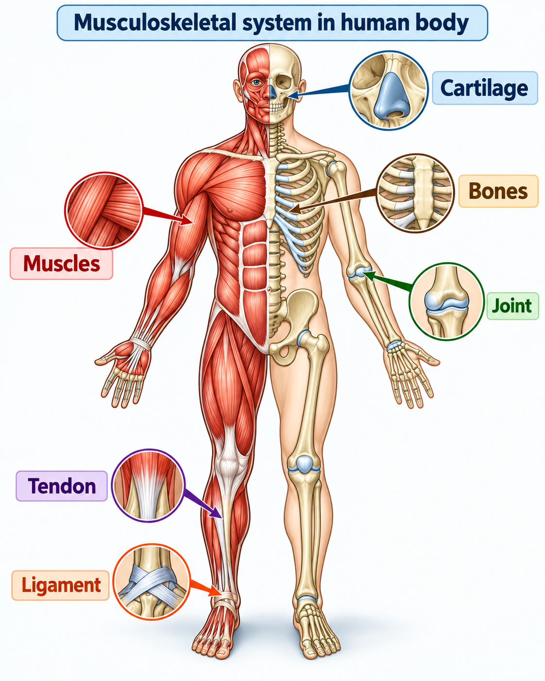
The musculoskeletal system consists of bones, muscles, joints, cartilage, tendons, and ligaments. Together, these structures provide support to the body, help us stand upright, move, and maintain posture, and protect important internal organs. In adults, the skeleton accounts for about 12–15% of total body weight.
Components of the Musculoskeletal System
The main components of the musculoskeletal system are:
- Bones – form the framework of the body, support body weight, protect internal organs, and help in movement.
- Muscles – contract and relax to produce movement and maintain posture.
- Joints – places where two or more bones meet; they allow different types of movement.
- Cartilage – a smooth, flexible tissue that cushions joints and reduces friction between bones.
- Tendons – strong bands of connective tissue that connect muscles to bones.
- Ligaments – tough bands of connective tissue that connect bones to bones and help stabilise joints.
Types of Joints
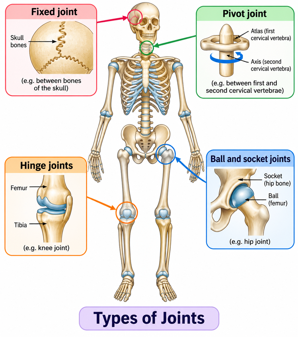
- Ball-and-socket joint: In this type of joint, the rounded end of one bone fits into a cup-like socket of another bone. It allows movement in many directions, including rotation. The shoulder and hip are examples of ball-and-socket joints.
- Hinge joint: A hinge joint allows movement mainly in one direction, similar to the opening and closing of a door. The elbow and knee are common examples. In the knee, the kneecap (patella) helps protect the joint.
- Pivot joint: A pivot joint allows one bone to rotate around another. In the neck, the first two cervical vertebrae form a pivot joint that helps the head turn from side to side, such as when we shake our head to say “no.”
- Fixed joint: Fixed joints do not allow movement between the bones. The bones of the skull are connected by fixed joints. These strong connections help protect delicate organs such as the brain, eyes, and ears.
course
No courses found
What are Tissues in Action Class 9 Notes?
Tissues in Action Class 9 Notes are study notes covering the important concepts of Class 9 Science Chapter 3. Infinity Learn provides these notes in a simple, student-friendly format with explanations, important points, tables, and diagrams for easier revision.
What topics are covered in Tissues in Action Class 9 Notes?
The notes cover plant tissues, meristematic tissues, permanent tissues, xylem, phloem, animal tissues, epithelial tissue, connective tissue, muscular tissue, nervous tissue, the musculoskeletal system, joints, totipotency, and crown gall disease. Students can study these topics with the help of Infinity Learn notes.
Are diagrams included in Tissues in Action Class 9 Notes?
Yes. The Infinity Learn notes include relevant diagrams and visual explanations to help students understand concepts such as meristematic tissues, xylem and phloem, animal tissues, neurons, muscles, joints, and totipotency more easily.
Can I download the Tissues in Action Class 9 Notes PDF?
Yes, students can download the Tissues in Action Class 9 Notes PDF from Infinity Learn and use it for offline study, quick revision, homework, and examination preparation.
How can Tissues in Action Class 9 Notes help in exam preparation?
The Infinity Learn notes provide concise explanations, important definitions, comparison tables, diagrams, and key revision points. Students can read the detailed explanations first and then use the PDF for quick revision before tests and exams.
