
Table of Contents
Structure Of The Human Heart: The heart is an organ that serves as a pump to circulate blood. It may be a consecutive tube, as in spiders and annelid worms, or a somewhat more elaborate structure with one or more receiving chambers (atria) and the main pumping chamber (ventricle), as in mollusks. The heart is a folded tube with three or four enlarged areas corresponding to fish’s mammalian heart chambers. In creatures with lungs—amphibians, reptiles, birds, and mammals—the heart shows various stages of evolution from a single to a double pump that circulates blood (1) to the lungs and (2) to the body as a whole. In humans, other mammals, and birds, the heart is a four-chambered double pump at the center of the circulatory system. In humans, the heart is situated between two lungs and slightly to the left of the center, behind the breastbone; it rests on the diaphragm, the muscular partition between the chest and the abdominal cavity.
Overview


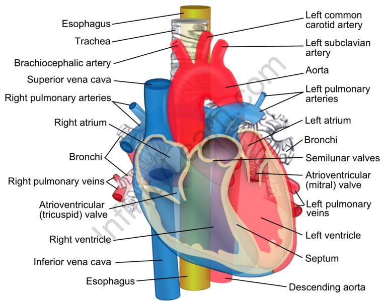
The heart consists of several layers of a formidable muscular wall, the myocardium. A pale layer of tissue, the pericardium, wraps the outside, and another layer, the endocardium, lines the inside. The heart cavity is divided down the inner into a right and a left heart, which in turn are subdivided into two chambers. The upper chamber is called an atrium (or auricle), and the lower section is called a ventricle. The two atria act as receiving rooms for blood entering the heart; the more muscular ventricles pump the blood out of the heart. Although a single organ, the heart can be considered two pumps that propel blood through two different circuits. The right atrium receives venous blood from the head, chest, and arms via the large vein called the superior vena cava and blood from the abdomen, pelvic region, and legs via the inferior vena cava. Blood then passes through the tricuspid valve to the right ventricle, propelling it through the pulmonary artery to the lungs. Venous blood connects with inhaled air, picks up oxygen, and loses carbon dioxide. Oxygenated blood is reimbursed to the left atrium through the pulmonary veins. Valves in the heart allow blood to flow one way and help maintain the pressure required to pump the blood.
Structure of the human heart
The human heart is about the size of a human fist and is divided into four chambers: two ventricles and two atria. The ventricles are the chambers that pump blood, and the atrium is the chambers that receive blood. The right atrium and ventricle make up the “right heart,” and the left atrium and ventricle make up the “left heart.” The heart’s structure also houses the most prominent artery in the body – the aorta.
A wall of muscle named the septum separates the heart’s right and left parts.


Classification of the human heart
The human heart is positioned to the left of the chest and is enclosed within a fluid-filled cavity described as the pericardial cavity. The pericardial cavity walls and lining comprise a membrane known as the pericardium.
The pericardium is a fiber membrane found as an external covering around the heart. It protects the heart by producing a serous fluid, which lubricates the heart and prevents friction between the surrounding organs. The pericardium has two exclusive layers—
- Visceral Layer: It directly covers the outside of the heart.
- Parietal Layer: It forms a sac around the outer region of the heart that contains the fluid in the pericardial cavity.
Structure of the Heart Wall
The heart wall is made up of 3 layers, namely:
- Epicardium – Epicardium is the outermost layer of the heart. It is composed of a thin-layered membrane that lubricates and protects the outer section.
- Myocardium – This is a muscle tissue membrane that constitutes the heart’s middle layer wall. It contributes to the thickness and is responsible for the pumping action.
- The endocardium is the innermost layer that lines the inner heart chambers and covers the heart valves. Furthermore, it prevents the blood from sticking to the inner walls, preventing potentially fatal blood clots.
Internal Structure of Heart
The internal structure of the heart is rather intricate, with several chambers and valves that control the flow of blood.


Chambers of the Heart
Vertebrate hearts can be classified based on the number of chambers present. For instance, most fish have two rooms, and reptiles and amphibians have three sections. Avian and mammalian hearts consists of four areas. Humans are mammals; hence, we have four teams, namely:
- Left atrium
- Right atrium
- Left ventricle
- Right ventricle
Atria are thin, less muscular walls and are smaller than ventricles. These are the blood-receiving chambers that are fed by the large veins.
Ventricles are larger and more muscular chambers responsible for pumping and pushing blood out to circulation. These are connected to larger arteries that deliver blood for circulation.
The right ventricle and right atrium are comparatively smaller than the left chambers. The walls consist of fewer muscles than the left, and the size difference is based on their functions. The blood originating from the right side flows through the pulmonary circulation, while blood arising from the left chambers is pumped throughout the body.
Blood vessels
Blood vessels are the channels or conduits through which blood is distributed to body tissues. The vessels make up two closed systems of tubes that begin and end at the heart. The other system, the systemic vessels, carries blood from the left ventricle to the tissues in all body parts and returns the blood to the right atrium. Based on their structure and function, blood vessels are classified as either artery, capillaries, or veins based on their structure and function.
Classification of blood vessels
- Arteries
Arteries carry blood away from the heart—pulmonary arteries transport blood with low oxygen from the right ventricle to the lungs. Systemic arteries carry oxygenated blood from the left ventricle to the body tissues. Blood is pumped from the ventricles into large elastic arteries that repeatedly branch into smaller and smaller arteries until the branching results in tiny streets called arterioles. The arterioles play a crucial role in regulating blood flow into the tissue capillaries. About 10 percent of the total blood volume is in the systemic arterial system at any time. It supports the vessel and changes vessel diameter to regulate blood flow and pressure. The outermost layer that attaches the ship to the surrounding tissue is the tunica externa or tunica adventitia. This membrane is connective tissue with varying quantities of elastic and collagenous fibers. The connective tissue in this layer is quite dense and adjacent to the tunic media, but it changes to loose connective tissue near the vessel’s periphery.
- Capillaries
Capillaries, the most minor and most numerous of the blood vessels, form the connection between the vessels that carry blood away from the heart (arteries) and the vessels that return blood to the heart (veins). The primary purpose of capillaries is the exchange of materials between the blood and tissue cells.
- Veins
Veins carry blood toward the heart. After blood passes through the capillaries, it enters the minor veins, called venules. It flows into progressively larger and larger veins from the venules until it reaches the heart. This blood has an exacerbated oxygen content because it has just been oxygenated in the lungs. The walls of veins have the same three layers as the arteries. Although all the layers exist, there is short, smooth muscle and connective tissue. It makes the walls of veins thinner than streets, which is related to blood in the veins having less pressure than in the arteries. Because the walls of the veins are more delicate and less rigid than arteries, veins can hold more blood. Nearly 70 percent of the total blood volume is in the veins at any time. Medium and enormous seams have venous valves, similar to the semilunar valves associated with the heart, which help keep the blood flowing. Venous valves are essential in the arms and legs, where they prevent the backflow of blood in response to the pull of gravity.
Connotation of the chapter for JEE primary, NEET, and board exams
Studying the heart and its functions is essential because the heart’s function in any organism is to maintain a steady flow of blood throughout the body. It reloads oxygen and circulates nutrients among the cells and tissues.
- One of the essential functions of the human heart is to pump blood throughout the body.
- Blood delivers oxygen, hormones, glucose, and other elements to various body parts, including the human heart.
- The heart also substantiates that adequate blood pressure is maintained in the body.
Various structures of the human heart, including a detailed analysis of heart function and its importance in the human body, is available here. There are many materials and quantities in bio. Distinct units can be used to express different amounts in biology. Students who want to flourish in bio can get a good knowledge of specific topics from the article. The comprehensive unit of the human heart and its structure is provided here to assist students in effectively understanding the respective issue. Continue to visit our website for additional biology help.
Also read: Important Topic Of Biology: Cardiac Output.
FAQs
Define veins.
Veins carry blood toward the heart. After blood passes through the capillaries, it enters the minor veins, called venules.
What do you understand by blood vessels?
Blood vessels are the channels or conduits through which blood is distributed to body tissues.
Question: Explain the chambers of the heart.
Answer: Vertebrate hearts can be classified based on the number of chambers present. Avian and mammalian nuclei consist of four areas. Humans are mammals; hence, we have four teams, namely:
- Left atrium
- Right atrium
- Left ventricle
- Right ventricle

