Table of Contents

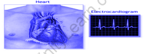
Electrocardiography is the process of creating an electrocardiogram (ECG or EKG), which is a recording of the electrical activity of the heart. It is a heart electrogram, which is a graph of voltage versus time of the electrical activity of the heart performed with electrodes placed on the skin.
During each cardiac cycle, these electrodes detect the small electrical changes caused by cardiac muscle depolarization and repolarization (heartbeat). Several cardiac abnormalities cause changes in the normal ECG pattern, including cardiac rhythm disturbances (such as atrial fibrillation and ventricular tachycardia, inadequate coronary artery blood flow (such as myocardial ischemia and myocardial infarction, and electrolyte disturbances (such as hypokalemia and hyperkalemia.
A healthy heart has an orderly progression of depolarization during each heartbeat that begins with pacemaker cells in the sinoatrial node, spreads throughout the atrium, and passes through the atrioventricular node down into the bundle of His and into the Purkinje fibres, spreading down and to the left throughout the ventricles.
The characteristic ECG tracing is caused by this orderly pattern of depolarization. An ECG provides a wealth of information about the structure of the heart and the function of its electrical conduction system to a trained clinician.
The overarching goal of an ECG is to obtain information about the electrical functioning of the heart. The medical applications of this information are diverse, and it is frequently required to be combined with knowledge of the structure of the heart and physical examination signs to be interpreted. The following are some indications for performing an ECG:
- Chest pain or a suspected myocardial infarction (heart attack), such as ST elevated myocardial infarction (STEMI)[16] or non-ST elevated myocardial infarction (non-STEMI (NSTEMI)
- Shortness of breath, murmurs, fainting, seizures, strange turns, or arrhythmias, including new-onset palpitations, or monitoring of known cardiac arrhythmias
- Medication monitoring (for example, drug-induced QT prolongation, Digoxin toxicity) and overdose management (e.g., tricyclic overdose), Hyperkalemia is an example of an electrolyte abnormality.
- Machines that consist of a set of electrodes connected to a central unit record electrocardiograms.
- Early ECG machines were built with analogue electronics, with the signal driving a motor that printed the signal on paper. Analogue-to-digital converters are now used in electrocardiographs to convert the electrical activity of the heart to a digital signal.
- Many ECG machines are now portable, with a screen, keyboard, and printer mounted on a small wheeled cart. Recent electrocardiography advancements include the development of even smaller devices for use in fitness trackers and smartwatches.
- These more compact devices frequently rely on only two electrodes to deliver a single lead I. There are also portable six-lead devices available.
FAQs
In physics, how does an electrocardiogram work?
Lead wires connect the electrodes to an ECG machine. The heart's electrical activity is then measured, interpreted, and printed. There is no electricity introduced into the body. Natural electrical impulses coordinate contractions of various parts of the heart to keep blood flowing normally.
What does an electrocardiogram mean?
An electrocardiogram (ECG or EKG) is a test that records the electrical signal from the heart in order to diagnose various heart conditions. Electrodes are placed on the chest to record the electrical signals produced by the heart, which cause the heart to beat.


