
Table of Contents
Cytoskeleton: A cytoskeleton is a type of temporary structure that exists in all cells within any living organism. It is made up of proteins and maintains cell structure, protects the cell, and enables all cells to move freely through certain structures such as flagella and cilia. It helps to move inside the cytoplasm like the movement of vesicles and organelles, for example, and aids the process involved in cell division.
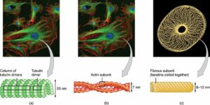
The cytoskeleton is responsible for shrinkage, cell mobility, movement of organelles and vesicles through the cytoplasm, cytokinesis, intracellular organisation of the cytoplasm, cell polarity formation, and many other important functions in cellular homeostasis and survival. It performs these functions with three basic structures: 7 to 8-nm wide microfilaments composed of actin, 10-nm IFs with cell-specific structure, and 24-nm external microtubules composed of -tubulin dimers. A cytoskeleton is a flexible structure in which three large fibers and tubes are under the influence of proteins that control their length, polymerization status, and degree of bonding. Myosin family members move vesicles near the actin microfilaments in a certain way, while family members of kinesin / KRP and dynein transport loads in smaller tracks and play a key role in the formation and function of the mitotic spindle. Both transfer types require ATP hydrolysis.
Cytoskeleton Definition
The cytoskeleton is a network of fibers and tubes that extend across the cell, through the cytoplasm, which is everything inside a cell except the nucleus itself. It is found in all cells, although the proteins they make from it vary from body to body. The cytoskeleton supports cells, shapes organelles, organizes and transforms them, and plays a role in molecular transport, cell division, and cell signalling.
Cytoskeleton structure and function
Eukaryotic cells are complex nucleus cells. There are eukaryotic cells in plants, animals, fungi, and protists. Prokaryotic cells are relatively simple, have no nuclei or true organelles other than ribosomes, and are found in bacteria and archaea of one living cell.
The cell cytoskeleton ensures stability, strength, and mobility. This provides the mobile scaffolding that organises the mobile organization. The figure represents part of the cytoskeleton of the cell. Note that the cytoskeleton is very large. Note that the cytoskeleton appears to have many ribosomes attached to it. Polysome refers to two ribosomes or more. The ribosomes attached to the cytoskeleton are often called free ‘ribosomes’ to separate them from the ribosomes attached to the nuclear membrane or ER.


Cytoskeleton structure
Three types of Cytoskeleton Components:
- Microfilaments
- Microtubules
- Middle threads
Microfilaments
Microfilament is the structure of the cytoskeleton filament, which includes actin monomers (f-actin). Here, globular g – actin monomers are polymerized to form strands of actin polymers (f-actin), more commonly known as g – actin. Finally, each filament (microfilament) is made up of two f-actin coils that have been misplaced.
It has also been shown that microfilament fibers have both positive and negative endings that contribute to the control of filaments in both ends. Studies have found, with respect to the development of microfilaments, that new monomers appear to be introduced at a faster rate compared to a bad end in a good end. There is also a limit to ATP in this good storage, which helps balance during rapid growth.
Microfilaments The thinnest / smallest microfilaments have a diameter of between 3 and 5 nm in diameter compared to other parts of the cytoskeleton. However, as actin compounds, microfilaments are rapidly accumulated and contribute to the proper functioning of the cell.
Microfilaments are usually found in the surrounding area of the cell, where they run from the plasma membrane to the microvilli (e.g. can be found in the pericanalicular area where they form pericanalicular web/meshwork). Here are the masses that form the intracellular – three-dimensional meshwork.
Microfilaments are very diverse and diverse although they are very small components of cytoskeletons.
Microtubules
Microtubules are the largest of the three parts of the cytoskeleton, with a diameter of 15 to 20 nm. It, unlike microfilaments, consists of a single type of glucose protein known as tubulin (a protein composed of kd polypeptides and alpha and beta-tubulin).
In favourable conditions, tubulin heterodimers join within the cell to form specific protofilaments. Such fibres combine to form microtubules (empty straws that resemble tubes).
Like microfilaments, cells tend to organize microtubules into clusters. However, with certain microtubules passing through the growth cycles and shortening their population, they are also shown to be less stable.
Heterodimer subunits are separated by specific tube ends during the reduction processes but are included during the growth phase. This complex instability has been caused by high internal variability and microtubule size.
Each microtubule contains approximately 13 protofilament lines arranged around an empty spine.
In a cell, microtubules appear in the form of a hub from the center of a cell. They emerge from the cytoplasm where they perform several functions.
Intermediate Fibers / Intermediate Filaments (IF)
Unlike other elements of cytoskeletons, the intermediate filaments comprise a large family of polypeptides. For this reason, different types of cells contain many different types of intermediate fibers.
Studies have shown that there are more than 50 different types of intermediate filaments divided into six major groups including:
- Type 1 and II – In most epithelial cells, they contain about 15 different proteins.
- Type III – This group of proteins binds like vimentin and desmin.
- Type IV – This group includes α-internexin-like proteins and neurofilament proteins found in nerve cells.
- Type V – Lamin is an example of a protein found in that group.
- Type VI – It is found in neurons, such as nestin.
Keratin is one of the most important proteins involved in the formation of intermediate fibres. This is a protein commonly found in the skin and hair.
The base rods between the two polypeptide chains are first twisted during bonding to form a folded structure (dimer). The outgoing dimers then combine to form tetramers that combine to form protofilaments at their ends (end to end). Finally, protofilaments are assembled to form intermediate fibres.
Depending on the size, the central fibres range from 8 to 10 nm in diameter — hence the term “medium fibres.” They are similarly stronger than the other two and are therefore more enduring.
Although they do not experience variable instability, as with microtubules, proteins are often converted by phosphorylation from intermediate filaments. This plays an important role in their integration within the cell.
The central fibres of a variety of cells extend from the nucleus to the cell membrane. Such fibres also connect with other parts of the cytoskeleton through a complex network that builds up in the cytoplasm, contributing to their functions.


Cytoskeleton function
There are several functions in the cytoskeleton. Second, it gives the shape of a cell. This is especially important for cells that do not have cell walls, such as animal cells, that do not get their shape from the outer layer. It can provide cell movement, too. Microfilaments and microtubules can disperse, reassemble and melt, allow cells to crawl and migrate, and microtubules can help build structures like cilia and flagella that facilitate cell movement.
The cytoskeleton organizes the cell and holds cell organelles in place, but it also helps move organelles throughout the cell. For example, microfilaments attract a vesicle that contains particles rolled into a cell during endocytosis when a cell inserts a molecule. Similarly, during cell division, the cytoskeleton helps move chromosomes.
The foundation of the house refers to the cytoskeleton. Like a house frame, the cytoskeleton is the “core” of the cell, which holds structures in place, provides support, and gives the cell a specific shape.
