Table of Contents
Definition:
The eye system is comprehended in three covers or coats, within which it consists of three other transparent coats. The uppermost structure consists of the cornea or sclera. In the center, we have a vascular tunic, or we can reach it uvea, and it is brief of the choroid, ciliary body and in this iris is also there. Further, the retina’s innermost area circulation from the choroid and retinal vessels. The aqueous humor, the ethical body, and adjustable lens within these covers. The function of the eye is elementary to understand as when the light is reflected off a surface and enters into the eye, cornea; vision begins, which refract rays through the pupil.
The ear’s structure can be outer, inner, and middle parts. The visible part of the ear is the outer part consisting of an article or pinna, and it channels the sound waves into the ear canal where it gets amplified and from where the waves travel towards a membrane that vibrates. After that, sound enters the inner part of the ear, but before this, in the middle part of the ear, the vibrations set the cochlea, which is filled with the fluid and moves when vibrated same as cochlea as it is a fluid also which moves when jiggled. After all this, the nerves are set into motions that further become electrical impulses and travel to the brain, where it is interpreted.
Overview
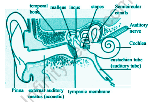
Structures of the human ear. The cartilaginous auricle and, therefore, the auditory meatus of the external ear direct sound waves to the center ear. The eardrum, stretched across the end of the channel, vibrates as sound waves reach it. Vibrations are transmitted via three tiny bones (hammer, anvil, stirrup) to the membranous oval window, linking the middle ear to the inner ear. The cochlea may be a coiled, fluid-filled tube lined with sensory hairs. Vibrations in the elliptic window cause movement of the cochlear fluid, stimulating the follicles to initiate impulses that travel along a branch of the auditory nerve to the brain. The eustachian tube, running from the middle ear to the nasopharynx, equalizes pressure between the center and outer ear. The fluid-filled semicircular canals play a task in balance, as hairs within the trenches answer movement-induced changes within the fluid by initiating impulses that visit the brain.
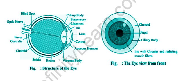
Structure of the human eye. The outer surface of the portion comprises the white protective sclera and transparent cornea, through which light enters. The middle layer encompasses the blood-supplying choroid and pigmented iris. Light passing into the inside through the pupil is regulated by muscles that control the pupil’s size. The retina comprises the third layer and contains receptor cells (rods and cones) that transform light waves into nerve impulses. The lens, fabricating directly behind the iris, focuses light onto the retina. Within the center of the retina, the macula may be a region of high acuity and color discrimination. Nerve fibers pass through the optic nerve to the brain’s optical center. The vitreous humor helps maintain the eye’s shape. A thin layer of mucosa (conjunctiva) protects the eye’s exposed surface. External muscles, including the medial rectus and lateral rectus muscles, connect and move the watch in its socket.
Elementary structure and function of eye and ear

Structure of Eye
The structure of an eye comprises three coats, within which further are three transparent structures. The outermost layer of the fibrous tunic consists of the cornea and sclera. We have the vascular tunic or uvea in the middle layer, consisting of the choroid, ciliary body, and iris. Moving on to the innermost layer is the retina. It acquires its circulation from the vessels of the choroid and also from the retinal vessels.
The flexible lens, aqueous humor, and the vitreous body lie within these coats. Vision begins when light is reflected off a surface and enters the eye through the cornea, refracting the rays through the pupil. The light rays then pass through the lens, which changes shape, bending the rays further and focusing on the retina.
Common disorders caused in Eyes
The most anticipated vision problems are diplopia ( hypermetropia), presbyopia ( vision), diplopia (age-related vision), and presbyopia. Presbyopia goods when the eye’s curve is not genuinely spherical, so light is concentrated inversely. Diplopia and presbyopia do when the vision is too narrow or broad to focus on the retina. The focal point is before the retina; insight it’s past the retina. In diplopia, the lens is strengthened, so it’s hard to bring close objects into focus.
Other eye problems include glaucoma ( increased fluid pressure, which can damage the optical whim-whams), cataracts (clouding and hardening of the lens), and macular degeneration ( degeneration of the retina).
Some facts about Eyes
The functioning of the eye is relatively simple, but there are some details you might not know:
- The eye acts precisely like a camera because the image formed on the retina is inverted (upside down). When the brain translates the picture, it automatically flips it. If you wear special goggles that make you view everything upside down, after a few days, your brain will adapt, again showing you the “correct” view.
- The lens soaks it before it can reach the retina. The reason humans evolved not to see UV light is that the light has enough energy to damage the rods and cones. Insects perceive ultraviolet light, but their compound eyes don’t focus as sharply as human eyes, so the point is spread over a larger area.
- Visionless people who still have eyes can feel the disparity between light and dark. There are special cells in the eyes that detect light but aren’t forming images.
- Each eye has a small blind spot. It is the moment where the optic nerve attaches to the eyeball. The hole in vision isn’t apparent because each eye fills in the other’s blind spot.
- Doctors are unable to transplant an entire eye. The reason is that it’s too hard to reconnect the million-plus nerve fibers of the optic nerve.
- Babies are born with full-size eyes. Human eyes remain nearly the same size from birth until death.
- Blue eyes contain no blue pigment. The color is a result of Rayleigh scattering, which is also responsible for the blue color of the sky.
- Eye color can change over time, mainly due to hormonal changes or chemical reactions in the body.

Structure of Ear
The ear’s structure can be broken down into three parts: the outer, inner, and middle. The outer ear consists of the auricle or pinna, the visible portion. It channels the sound waves into the ear canal, amplified by the waves traveling towards a vibrating membrane. In the middle ear, the pulses set the ossicles into motion. These sound waves enter the inner ear and then into the cochlea, filled with a fluid that moves with the beats.
Common diseases caused in ears
- Otitis media: an inflammation of the internal ear caused by a bacteria or virus that causes fluid to accumulate behind the eardrum. It’s not severe if treated correctly and doesn’t return repeatedly.
- Otosclerosis: the abnormal growth of the tiny bones within the tympanic cavity. It’s one of the foremost common causes of gradual deafness in adults, but surgery can recover hearing.
- Tinnitus: a sensation of noise within the head, like a continuing ringing or buzzing. There’s currently no scientific treatment or cure for this condition.
- Presbycusis is age-related deafness, the gradual loss of hearing in adults as they get older. It’s usually bilateral and symmetrical, meaning it occurs at an equivalent rate simultaneously in both ears.
- Barotrauma is the term for physical damage to the ear caused by barometric (air) or water pressure changes.
- Acoustic trauma is damage caused to the ear by a sudden bang like explosions, loud machinery, or music concerts. The impact and effects of the injury got to be assessed and monitored over the medium and future.
- We hear loss, impairment, or anacusis (deafness): the problem of listening thanks to partial, unilateral, bilateral, or total deafness. It’s going to be hereditary or the consequence of an illness, traumatism, long-term exposure to noise, or aggressive medication for the acoustic nerve. Hearing aids or cochlear implants are often wont to correct the hearing disorder.
Also read: Important Topic Of Biology: Thyroid
FAQs
Define the structure of human eyes.
The eye system is comprehended in three covers or coats, within which it consists of three other transparent coats. The uppermost structure consists of the cornea or sclera. In the center, we have a vascular tunic, or we can reach it uvea, and it is brief of the choroid, ciliary body and in this iris is also there. Further, the retina's innermost area circulation from the choroid and retinal vessels. The aqueous humor, the ethical body, and adjustable lens within these covers
How is the human ear structured?
The ear's structure can be broken down into three parts: the outer, inner, and middle. The outer ear consists of the auricle or pinna, the visible portion. It channels the sound waves into the ear canal, amplified by the waves traveling towards a vibrating membrane. In the middle ear, the pulses set the ossicles into motion. These sound waves enter the inner ear and then into the cochlea, filled with a fluid that moves with the beats.
What do you understand by the term myopia?
Condition during which you'll see objects almost you clearly, but things farther away are blurry. It occurs when the form of your eye causes light rays to bend (refract) incorrectly, focusing images ahead of your retina rather than on your retina.
Which disease is caused in human ears due to age?
Presbycusis is age-related deafness, the gradual loss of hearing in adults as they get older. It's usually bilateral and symmetrical, meaning it occurs at an equivalent rate simultaneously in both ears.


