Table of Contents

INTRODUCTION
The cell is considered a structural and functional unit of life. Each cell in the body is sealed with a bubble-like structure called a cell membrane. Also called a plasma membrane. Its main function is to separate the inside and the outside of the cell.
When discussing the structure of a cell membrane it is important to know what the cell membrane is made of? The cell membrane or plasma membrane not only helps to separate cell boundaries but also allows the cell to interact with the environment in a controlled and disciplined manner. Cells must be able to extract or take or extract certain objects at a certain value. In addition, they are able to communicate with other cells, by identifying other cells and sharing relevant information.
Here, we will look at the different parts of the plasma membrane. Cell membrane activity, membrane variation. The way these layers work together to form a sensitive, flexible, and secure boundary around the cell.
Plasma Membrane Structure
The accepted and currently used model to represent the formation of plasma membranes is called the liquid mosaic model. It was first proposed by S. J. Singer and G. L. Nicolson in 1972. The model was found to have evolved over time but is still able to provide a good basic definition of membrane formation in many cells.
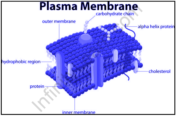
According to the fluid mosaic model structure, plasma membranes look like a mosaic with key components such as phospholipids, cholesterol, and proteins. These parts are found to move freely and without water in the lining of the membrane. Interestingly, this liquid can be described as inserting or piercing the membrane with a very fine needle. The membrane will simply separate to flow closer to the needle and once the needle has been removed, it returns to flow together without stitching.
Parts of the Plasma Membrane
The cell membrane is made up of lipids, proteins, and carbohydrate groups that attach to other lipids and proteins. Lipids include phospholipids and cholesterol.
The phospholipid is a type of lipid composed of glycerol, which has two fatty acids and a phosphate-attached head group. The biological membrane usually consists of two layers of phospholipids with their tails pointing inwards. This type of formulation is called phospholipid bilayer.
The cholesterol layer is another lipid layer composed of four carbon-bound rings. It is found on the sides of the phospholipid layer in the center of the membrane.
Membrane proteins can extend the path inside the plasma membrane, where these proteins are able to cross the membrane completely or can be freely connected to their inner or outer surface.
Carbohydrate groups are found only outside the plasma membrane. It is attached to proteins to form glycoproteins, or lipids to form glycolipids.
The levels of lipids, proteins, and carbohydrates present in the plasma membrane may vary from one cell to another. In a normal cell, proteins can contribute up to 50 percent of the total amount. Although, lipids of all kinds contribute about 40 percent. The remaining 10 percent are given carbohydrates.
Cell Function
The cell membrane acts as a barrier between two different regions and the gatekeepers. They are able to penetrate slowly naturally, which means that fewer molecules are allowed to spread throughout the lipid bilayer. Small hydrophobic cells and gases such as oxygen and carbon dioxide can cross the membrane very quickly. Small tropical molecules, such as water and ethanol, may pass through the membrane, but may penetrate slowly. The cell membrane can limit the process of dispersing highly charged molecules and large molecules. Highly charged molecules contain ions and larger molecules contain sugar and amino acids. The passage of these molecules depends entirely on the transport of certain proteins embedded in the membrane.
The transport proteins in the membrane are very clear and selective that can travel across the membrane and often use energy to make the passage. Also, proteins transport certain nutrients against concentration gradients. This also imposes an additional requirement for additional power. The ability of the membrane to maintain concentration gradient and move materials against them is essential for cell life and maintenance.
Other types of transmembrane proteins are involved in communication-related activities. These types of proteins can bind to signals, such as hormones or body coordinates. The binding of these proteins can change the protein that can transmit signals to other atoms within the intracellular messenger. Like transport proteins, receptor proteins are also active and selective in nature.
Membrane Cell Variation
In contrast to single-celled cells or prokaryotes, eukaryotic cells and plasma membranes, also have intracellular membranes that can surround various organelle. In these cells, the plasma membrane is part of a broader endometrial system. The system includes (ER) endoplasmic reticulum, nuclear membrane, Golgi apparatus, and lysosomes.
Membrane components are systematically exchanged throughout the endomembrane system. For example, the ER and the Golgi membrane have different combinations. The proteins found in these membranes include filter signals, which act as cellular zip codes that can determine their final location.
Mitochondria and chloroplasts are also surrounded by membranes, but they have unusual membrane structures. Each of these organelles instead of having one membrane becomes two surrounding membranes. The outer layer of these organelles has holes that allow for the passage of small molecules. The inner membrane is packed with proteins that make up the electron transport chain. These membrane layers of mitochondria and chloroplasts are similar to other prokaryotes today.
Membranes are composed of lipids and proteins, which act as a barrier to the specific functions of cells and organelles within the cell. These membranes can store external molecules “outside” and internal molecules “inside”. This allows certain molecules to transmit messages through a series of cellular events.
Introduction of Endoplasmic Reticulum
The endoplasmic reticulum (ER) is a large organelle made up of membranous sheets and tubes that begin near the nucleus and extend throughout the cell. The endoplasmic reticulum produces, binds, and secretes many cellular products.
Definition
What is the Endoplasmic Reticulum?
The Endoplasmic reticulum (ER) is one of the most important organelles in the cell. Although the function of the nucleus is to function as a brain cell, the ER acts as a production and packaging system. Golgi materials, ribosomes, mRNA, and tRNA work closely with the ER.
The Endoplasmic Reticulum Structure is a network of cells that are distributed to the cell and connected to the nucleus. The membrane varies from cell to cell, and ER size and shape are determined by cell function.
For example, prokaryotes or red blood cells do not have ER of any kind. Cells binding and releasing many proteins will require a large amount of ER. For good examples of cells with large ER structures, you may look at a cell from the pancreas or liver.
Before going into the details, first, check out the drawing of the endoplasmic reticulum:
Endoplasmic Reticulum formation
The ER membrane system can naturally be divided into two structures – cisternae and sheets.
The Cisternae are structurally tubular and form a three-dimensional polygonal-dimensional network. In mammals, they are about 50 nm wide, and yeast, about 30 nm wide.
On the other hand, ER sheets are membrane-closed, flat-sized sacs, which extend across the cytoplasm. They are often associated with ribosomes and special proteins called translocons, which are needed to translate proteins within RER.
Types of Endoplasmic Reticulum
The endoplasmic reticulum (ER) provides about 50 percent of the total membrane over the animal cell. Organelle called ‘endoplasmic reticulum’ is present in plants and animals and is an important source of lipids (fats) and other proteins. Some of these materials are made for other organelles and are exported to them.
The endoplasmic reticulum is divided into two types: the endoplasmic reticulum and the smooth endoplasmic reticulum.
In both animal and plant cells, the endoplasmic reticulum is visible.
Cells that specialize in protein production will continue to have heavy ER, while cells containing lipids (fat) and steroid hormones will have a smooth ER.
- Rough Endoplasmic Reticulum
Rough ER (RER) is also listed in the ribosomes section and is very important for protein synthesis and packaging.
Ribosomes are attached to the ER membrane, making it “hard”. Also, RER is attached to a nuclear envelope around the nucleus. This direct correlation between the perinuclear space and the ER lumen causes molecules to pass through both membranes. The process of protein synthesis begins when mRNA passes from the nucleus to the ribosome in the RER surface.
As the ribosome forms a chain of amino acids, the chain is forced into the RER’s cisternal area. When protein production is complete and this is collected, RER presses the vesicle. The vesicle, a small bubble, can enter the cell membrane or into the Golgi machine. Some proteins are used in the cell and others are sent to the space between cells.
- Smooth Endoplasmic Reticulum
The smooth endoplasmic reticulum (SER) acts as a storage organelle. It is essential for lipid and steroid formation and maintenance. Steroids are a type of ringed organic molecule, which is used in living things for many purposes. It is more than just the building of a weight lifter. Your body’s fat-releasing cells also have more SER than most cells. A variant of the SER is the sarcoplasmic reticulum (SR). It can store a few ions in the solution that the cell will need over time. If a cell has to act fast, scanning the atmosphere for additional ions that may be floating or floating does not make sense. Therefore, it retains the ions needed for this process.
- Ultrastructure of Endoplasmic Reticulum
All three structures of the Endoplasmic reticulum are surrounded by a thick layer. The membrane also consists of three layers – the outer and inner layers contain protein molecules, while the phospholipids are two thin and flexible medium layers. The endoplasmic reticulum membrane of the plasma membrane, the nuclear membrane, and the Golgi complex membrane continue. The Endoplasmic reticulum lumen acts as a secret product, and in it, Palade (1956) acquired the author’s granules.
Functions of Endoplasmic Reticulum
- It is primarily responsible for the transfer of other organelle proteins and other carbohydrates, including lysosomes, Golgi apparatus, plasma membrane, etc.
- They also provide a cellular response to a high-altitude rise.
- They assist in the formation of a nuclear membrane during cell division.
- They play a vital role in shaping the skeletal system.
- They play an important role in protein, lipid, glycogen, and other steroid compounds, such as cholesterol, progesterone, testosterone, etc.
Also read: Important Topic Of Biology: Cell Envelope
FAQs
What is a cell membrane?
A cell membrane, also called a plasma membrane, is found in all cells and divides the inside of the cell from the outside.
Q. Write about the functions of the plasma membrane.
Ans: Thus, the cell membrane has two following functions.
- It acts as a barrier to storing cell elements inside and sends unwanted substances out.
- It acts as a gateway to allow the transport of essential nutrients and waste products.


