Table of Contents
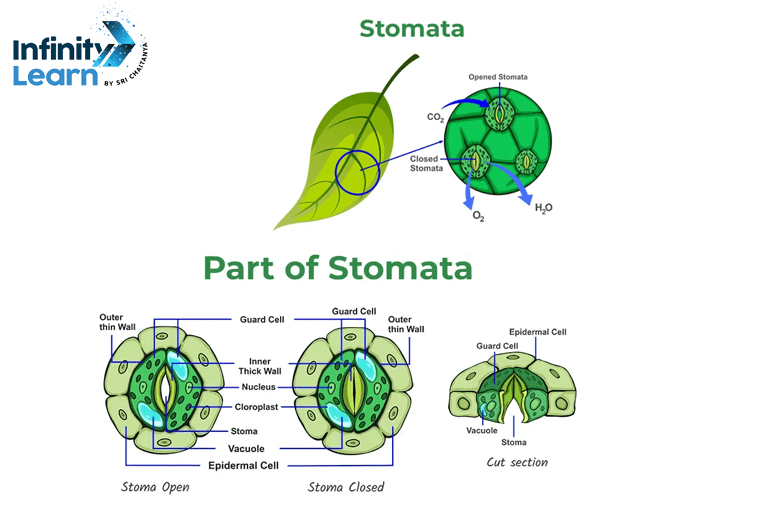
Stomata Diagram: All green plants have essential parts that play critical roles in various life processes. One key feature is the stomata, which are crucial for gaseous exchange. Plants use stomata as their mouths, also known as stoma or stomas.
Stomata typically exist in the epidermis of leaves, although they can also manifest on stems and other plant parts. These openings allow gases like oxygen, carbon dioxide, and water vapor to move between the inside and outside of plant tissues.
Stomata come in different types, classified based on the number and characteristics of the surrounding subsidiary cells. Below are some types of stomata. The diagram of stomata is important for students in both classes 10 and 12, frequently appearing in board exams. Below is a brief description and a well-labeled diagram of stomata for reference.
What is Stomata Diagram
A stomata diagram is a graphical representation showing the small openings, known as stomata, which are primarily located on the underside of plant leaves. These pores are surrounded by guard cells. They play a critical role in regulating the exchange of gases. This includes carbon dioxide and oxygen, as well as water vapor. This exchange occurs between the plant and its environment. The diagram helps in understanding the structure and function of stomata, including their involvement in processes like photosynthesis and transpiration.
Well-Labelled Stomata Diagram
Introducing a well-labelled diagram of open and closed stomata provides a clear understanding of these important plant structures. Below is a diagram of stomata, clearly labeling the different cells that make up its composition.

Structure of Stomata
The structure of stomata consists of tiny pores, primarily located in the epidermis of plant leaves, stems, and other organs. These pores are bordered by two specialized cells known as guard cells. These guard cells control the opening and closing of the stomatal pore, regulating the exchange of gases such as carbon dioxide, oxygen, and water vapor between the plant and its surroundings. Understanding the structure of stomata is essential for comprehending their function in plant physiology and gas exchange processes. A typical stomata comprises several distinct parts, each contributing to its function:
- Epidermal Cells
The outermost layer of a plant’s leaf consists of epidermal cells covered by a waxy cuticle. Beneath this layer lie the palisade cells containing chloroplasts, followed by the spongy tissue with air spaces. Epidermal cells serve to regulate transpiration and provide protection to the plant. - Subsidiary Cells
Subsidiary cells, also known as accessory cells, border the guard cells and support their function. These cells may aid in guard cell movement or act as reservoirs for ions and water, enhancing the efficiency of stomatal regulation. - Guard Cells
Guard cells, specialized kidney-shaped cells, flank the stomatal pore. They possess chloroplasts and feature a thin outer wall and a thick inner wall made of cellulose. Guard cells play a crucial role in regulating stomatal opening and closing, thereby controlling the exchange of gases between the leaf and the atmosphere. - Stomatal Pore
The stomatal pore is a microscopic opening on the surface of plant tissues, facilitating various physiological processes such as photosynthesis, water transport, and temperature regulation. During the day, these pores open to allow the entry of gaseous carbon dioxide for photosynthesis, while waste oxygen produced by photosynthesis exits through the same openings. Additionally, water vapor is released through stomatal pores in a process known as transpiration.
Stomata Diagram – Types of Stomata
Stomata can be classified into five types based on the arrangement of subsidiary cells surrounding the guard cells:
- Anomocytic Stomata: Irregularly shaped guard cells with no specific subsidiary cell arrangement.
- Anisocytic Stomata: Guard cells accompanied by three subsidiary cells of varying sizes.
- Paracytic Stomata: Paired guard cells with subsidiary cells aligned parallel to each other.
- Diacytic Stomata: Paired guard cells with subsidiary cells positioned at an angle to each other.
- Gramineous Stomata: Guard cells surrounded by subsidiary cells arranged in a parallel manner, characteristic of grasses and cereals.
Why Study Stomata Diagrams?
- Understand Plant Breathing: Stomata diagrams elucidate how plants respire by facilitating gas exchange, vital for photosynthesis and respiration.
- Explore Environmental Adaptation: Examination of stomata diagrams reveals how plants adapt to varying conditions like light, humidity, and carbon dioxide levels, optimizing their physiological processes.
- Insight into Plant Responses: By studying stomata diagrams, one gains insight into how plants respond to environmental changes, aiding in research across botany, agriculture, and environmental science.
- Enhance Crop Development: Knowledge of stomata structures informs the development of crop varieties with improved water use efficiency, contributing to agricultural sustainability.
- Fundamental Plant Science: Understanding stomata diagrams is foundational in plant science education, providing essential knowledge for botanists, researchers, and students alike.
Stomata Diagram FAQs
What is a stomata and how is it represented in a diagram?
Stomata, tiny pores on plant surfaces, help in gas exchange. Depicted in diagrams as small openings, they're surrounded by specialized cells like guard cells and subsidiary cells, highlighting their structure and function.
How do stomata open and close, particularly in Class 9?
Stomata open and close through the expansion and contraction of guard cells, which regulate their aperture. When guard cells absorb water, they swell and curve, causing the stomatal pore to open. Conversely, loss of water leads to shrinkage and straightening of guard cells, resulting in stomatal closure. This mechanism helps regulate gas exchange and minimize water loss in plants.
What is meant by the stomatal apparatus, particularly in Class 11?
The stomatal apparatus comprises stomata and specialized cells like guard cells, subsidiary cells, and epidermal cells. This arrangement regulates gas exchange (like CO2 and O2) and controls water vapor and transpiration in plants.
Can you draw a well-labeled diagram of stomata?
In the above article Infinity learn has provided a diagram with labels indicating key components such as guard cells, subsidiary cells, and stomatal pore, demonstrating the structure of stomata.


