Cell Biology for NEET: Simplest Guide to Cell Structure & Functions
By Karan Singh Bisht
|
Updated on 10 Jul 2026, 17:19 IST
Cell Biology forms the foundation of NEET Biology. Master this single unit, and topics like Genetics, Biotechnology, and Physiology become much easier to grasp. This segment reliably yields 6 to 8 questions every year, meaning you can secure up to 32 marks by mastering it.
This guide breaks down the core concepts exactly as explained in NCERT, highlighting where students usually lose marks.
Difference Between Prokaryotic and Eukaryotic Cells
The primary divide between prokaryotic and eukaryotic cells rests on how they manage genetic material and whether they contain internal membrane-bound compartments.
The Structural Layout
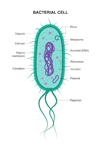
- Prokaryotes: Simpler, single-celled organisms lacking a well-defined nucleus. Their genetic material lies naked in the cytoplasm within an irregular region called the nucleoid.
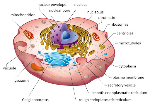
- Eukaryotes: Possess an organized internal structure. Their genetic material is enclosed within a double-layered nuclear membrane, and they contain specialized, membrane-enclosed organelles.
Key Divergences Explained
The differences between these two groups extend down to ribosome types and cell wall compositions. Let us look at these features side by side.
| Feature | Prokaryotic Cells | Eukaryotic Cells |
| Nuclear Status | Absent: no true nuclear membrane or nucleolus. | Present: distinct nuclear envelope enclosing genomic DNA. |
| Ribosome Type | 70S type: split into 50S and 30S subunits. | 80S type in cytoplasm: 70S inside organelles like mitochondria. |
| Cell Wall Chemistry | Composed of peptidoglycan in bacteria. | Cellulose in plants, chitin in fungi, absent in animals. |
| Genetic Material | Circular, single double-stranded DNA molecule without histones. | Linear, multiple double-stranded chromosomes wound around histones. |
| Membrane-bound Organelles | Completely absent. | Present: includes ER, Golgi bodies, lysosomes, plastids. |
| Examples | Bacteria, Cyanobacteria (blue-green algae), Mycoplasma. | Plant cells, Animal cells, Fungal cells, Protists. |
Real-World Taxonomic Examples
When tracking prokaryotes for your exam, focus heavily on Mycoplasma. Students often forget that these unique organisms lack a cell wall entirely. They are the smallest known living cells and can survive without oxygen. They frequently feature in matching-type questions in NEET.
Eukaryotic structure is visible across macroscopic life. In plant cells, you will find rigid cellulose walls and large central vacuoles. Animal cells omit the wall entirely, opting for an outer plasma membrane and internal centrioles to coordinate cell division.

Cell Organelles and Their Functions
Think of a eukaryotic cell as a busy factory. Every organelle is a dedicated department running a specific, non-interfering chemical process.
Organelles Found in BOTH Plant and Human Cells
- Nucleus: The cell command center. It is wrapped in a double-layered nuclear envelope perforated by microscopic nuclear pores. These pores regulate the movement of RNA and proteins between the nucleus and the cytoplasm. Inside, you will find chromatin (DNA wrapped around basic histone proteins) and the nucleolus, which is the site for active ribosomal RNA (rRNA) synthesis.
Remember for NEET: Cells that are actively producing proteins contain larger and more numerous nucleoli.
Best Courses for You

JEE

NEET

Foundation JEE

Foundation NEET

CBSE
- Mitochondria: The powerhouse of the cell. It generates cellular energy as ATP. It features a smooth outer membrane and a deeply folded inner membrane. These folds, called cristae, increase the surface area available for chemical reactions. The internal gel-like space is called the matrix.
Crucial NEET Fact: The mitochondrial matrix contains a single, circular, naked DNA molecule, a few RNA molecules, and 70S ribosomes. This allows mitochondria to synthesize some of their own proteins, making them semi-autonomous organelles.
- Endoplasmic Reticulum (ER): An expansive network of tiny tubular structures scattered throughout the cytoplasm.
- Rough Endoplasmic Reticulum (RER): Studded with ribosomes on its outer surface. It handles protein synthesis and routing.
- Smooth Endoplasmic Reticulum (SER): Lacks attached ribosomes. It specializes in lipid biosynthesis. In animal cells, the SER is the primary site where steroid hormones are manufactured.
- Golgi Apparatus: Consists of flat, disc-shaped sacs called cisternae stacked parallel to each other. The organelle is polarized, featuring a convex cis face (forming face) and a concave trans face (maturing face). It modifies, packages, and processes proteins coming from the RER into glycoproteins and glycolipids.
- Lysosomes: Spherical, membrane-bound vesicular structures formed by the packaging action of the Golgi apparatus. They are packed with hydrolytic enzymes (lipases, proteases, carbohydradates) optimized to function at an acidic pH.
- Ribosomes: Granular, non-membrane-bound structures composed of ribosomal RNAs and proteins. Discovered by George Palade, they serve as the site of protein synthesis in all living cells.
Organelles Found ONLY in Plant Cells (Absent in Human Cells)
- Chloroplast: Double-membrane, semi-autonomous organelles. The space limited by the inner membrane is called the stroma. Suspended within the stroma are flattened, membrane-bound sacs called thylakoids, which stack up like coins to form grana. The thylakoid membranes contain chlorophyll pigments to harvest light energy for photosynthesis.
- Large Central Vacuole: A massive space occupying up to 90% of a plant cell's volume. It is enclosed by a single, highly selective membrane called the tonoplast. The tonoplast actively pumps ions and other materials against concentration gradients into the vacuole, making the solute concentration significantly higher inside the vacuole than in the surrounding cytoplasm.
- Cell Wall: A rigid, non-living outer layer that provides structural shape and protects the cell from mechanical damage. Plant cell walls are primarily made of cellulose, hemicellulose, and pectins. Neighboring cell walls are glued together by a middle lamella composed largely of calcium pectinate.
Organelles Found ONLY in Animal/Human Cells (Absent in Plant Cells)
- Centrosome and Centrioles: The centrosome is an organelle usually containing two cylindrical structures called centrioles. They lie perpendicular to each other and form the basal body of cilia or flagella, and give rise to spindle fibers during cell division in animal cells. Plant cells manage cell division without centrioles.
Types of Transport Across the Cell Membrane
The plasma membrane is a dynamic, selectively permeable barrier. It regulates exactly what enters and exits the cell space.
The Fluid Mosaic Model
Singer and Nicolson proposed the Fluid Mosaic Model in 1972, which remains the widely accepted explanation for biological membranes. The membrane is organized as a lipid bilayer composed primarily of amphipathic phospholipids. Each phospholipid molecule possesses a hydrophilic (polar) head facing the watery exterior and two hydrophobic (non-polar) fatty acid tails hidden inside the interior.
Proteins are embedded within this lipid matrix like a mosaic. They are classified based on how easily they can be extracted from the membrane.
- Integral Proteins: These are deeply embedded or transit entirely through the bilayer. They serve as permanent structural channels or transport pumps.
- Peripheral Proteins: These lie on the surface of the membrane and can be easily detached.
The quasi-fluid nature of the lipid bilayer allows lateral movement of proteins within the membrane. This fluidity is essential for key cellular processes like cell growth, secretion, endocytosis, and cell division.
Molecular Transport Mechanisms
The plasma membrane is selectively permeable. Molecules cross this barrier using different mechanisms depending on their size, charge, and concentration gradients.
- Passive Transport: Movement occurs along a concentration gradient from an area of higher concentration to lower concentration without any expenditure of metabolic energy.
- Simple Diffusion: Neutral solutes move freely across the lipid bilayer along their gradient.
- Osmosis: The specific movement of water molecules across the selectively permeable membrane driven by an osmotic gradient.
- Facilitated Diffusion: Polar molecules and ions cannot pass through the non-polar lipid core on their own. They require specialized membrane proteins, known as carrier proteins or channel proteins, to facilitate their passage along the concentration gradient without consuming ATP.
- Active Transport: An energy-dependent process where specialized pump proteins transport molecules or ions against their concentration gradient, moving them from low to high concentration. This process requires direct ATP consumption. The sodium-potassium pump (Na^+/K^+ pump) is a classic example. It pumps three Na^+ ions out of the cell while pulling two K^+ ions in per ATP consumed.
- Bulk Transport: * Endocytosis: The cell membrane engulfs large particles or fluids, pulling them into the cytoplasm inside a vesicle.
- Exocytosis: Intracellular secretory vesicles fuse with the plasma membrane to expel their contents into the extracellular space.
Cell Cycle and Cell Division
A cell does not grow indefinitely. It undergoes a tightly regulated cycle of growth and division to maintain genetic continuity across generations. This process is divided into two basic phases: Interphase, where the cell prepares to divide, and the M Phase (Mitosis or Meiosis), where actual division occurs.
The Cell Cycle and Its Regulation
Students often skip the details of Interphase, but NEET papers frequently target this phase. Interphase takes up more than 95% of the total duration of the cell cycle.
- G1 Phase (Gap 1): The cell is metabolically active and continuously grows but does not replicate its DNA.
- S Phase (Synthesis): DNA replication occurs. Remember this rule for tracking chromosome numbers: the amount of DNA doubles (from 2C to 4C), but the total chromosome number remains exactly the same (2n). Centriole replication also begins in the cytoplasm of animal cells during this phase.
- G2 Phase (Gap 2): Proteins required for mitosis, like tubulin, are synthesized while cell growth continues.
- G0 Phase (Quiescent Stage): Some cells in adult animals do not divide, such as heart cells. These cells exit the G1 phase to enter an inactive metabolic state called the G0 phase. They remain metabolically active but no longer proliferate unless called upon by specific physiological cues.
The cell cycle is strictly monitored by internal regulatory checkpoints managed by proteins called Cyclins and Cyclin-Dependent Kinases (CDKs). If DNA damage is detected at the G1-to-S boundary or if spindle fibers fail to attach properly during the M phase, the cycle pauses to prevent incorrect chromosome distribution.
Mitosis: Equational Division
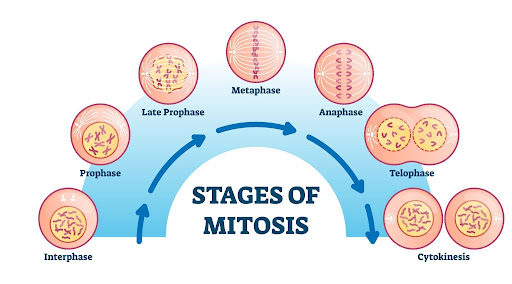
Mitosis occurs primarily in diploid somatic cells of animals and can occur in both haploid and diploid plant cells. It ensures that the daughter cells receive an exact duplicate of the parent cell's chromosome complement. It is divided into four sequential nuclear stages.
1. Prophase
Chromatin condensation occurs, making individual chromosomes visible under a microscope. Each chromosome is seen to consist of two sister chromatids attached at a central centromere. Centrosomes move to opposite poles and initiate spindle fiber assembly. The nucleolus, Golgi complexes, and nuclear envelope break down and disappear by the end of this phase.
2. Metaphase
The nuclear envelope is completely gone. Spindle fibers attach to the kinetochores, which are disc-shaped structures on the centromeres of each chromosome. Chromosomes align along the equator of the cell, forming the metaphase plate. This is the best stage to study chromosome morphology under a microscope.
3. Anaphase
The centromere of each chromosome splits simultaneously. Sister chromatids separate and are pulled toward opposite poles by the shortening of spindle fibers. Students often confuse chromatid numbers here, so remember that the chromosome number doubles temporarily during this phase because each separated chromatid is now considered an individual chromosome.
4. Telophase
Chromosomes reach their respective poles, decondense, and lose their individuality, reverting to a loose chromatin network. The nuclear envelope reforms around each chromosome cluster. The nucleolus, Golgi bodies, and ER reappear at both poles.
Following nuclear division (karyokinesis), the cytoplasm divides during cytokinesis. In animal cells, this happens via a contractile ring that forms a deepening furrow in the plasma membrane. Plant cells divide from the inside out by forming a cell plate, which eventually develops into the middle lamella.
Significance of Mitosis
- It maintains a constant chromosome number across somatic daughter cells.
- It is vital for growth, cell repair, and tissue regeneration (such as replacing old cells in the blood and gut lining).
Meiosis: Reductional Division
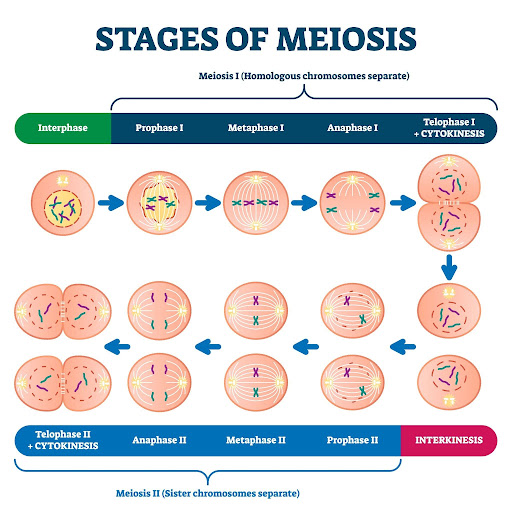
Meiosis occurs exclusively in specialized germ cells during gametogenesis. It reduces the chromosome number by half, converting a diploid cell (2n) into four genetically distinct haploid gametes (n). Meiosis consists of two consecutive nuclear divisions but only a single round of DNA replication.
Meiosis I (Reductional Division)
This stage separates homologous chromosomes, reducing the ploidy level from diploid to haploid. Prophase I is highly extended and subdivided into five distinct substages based on chromosomal behavior.
- Leptotene: Chromosomes become progressively visible under light microscopy as chromatin condensation begins.
- Zygotene: Homologous chromosomes begin pairing up side-by-side in a process called synapsis. This pairing is stabilized by a complex protein structure known as the synaptonemal complex. The paired chromosomes form a bivalent or tetrad.
- Pachytene: This is a critically important phase for NEET. Non-sister chromatids of homologous chromosomes undergo crossing over at specific points. The enzyme recombinase mediates this exchange of genetic material, creating new recombinant allele combinations that drive evolutionary variation.
- Diplotene: The synaptonemal complex dissolves. Homologous chromosomes begin to pull apart but remain glued at the exact points where crossing over occurred. These X-shaped contact points are called chiasmata. In the oocytes of certain vertebrates, diplotene can last for months or even years.
- Diakinesis: Chiasmata undergo terminalization as they slide off the ends of the chromosomes. The nucleolus degenerates, the nuclear envelope breaks down completely, and the meiotic spindle assembly prepares for homologous separation.
During Metaphase I, bivalent chromosomes align along the equatorial plate in two parallel lines. In Anaphase I, homologous chromosomes separate and move toward opposite poles while sister chromatids remain attached at their centromeres. Telophase I reforms the nuclear membrane, leading into a brief rest phase called interkinesis, during which no DNA replication occurs.
Meiosis II (Equational Division)
Meiosis II mimics a standard mitotic division. During Anaphase II, the centromeres split simultaneously, allowing sister chromatids to separate and move to opposite poles. The process concludes with four haploid daughter cells, each containing a unique genetic combination.
Significance of Meiosis
- It conserves the specific chromosome number of a species across generations by halving gamete ploidy.
- It introduces genetic variations due to crossing over, which are essential for evolution.
Key Conceptual Differences
To avoid confusion during fast-paced exams, keep the core differences between mitosis and meiosis clear.
| Property | Mitosis | Meiosis |
| Site of Occurrence | Somatic cells (growth and tissue repair). | Germ cells (gamete production). |
| Number of Divisions | Single nuclear and cytoplasmic division. | Two successive nuclear and cytoplasmic divisions. |
| Daughter Cell Count | Two diploid (2n) daughter cells. | Four haploid (n) daughter cells. |
| Genetic Identity | Identical twins of the parent cell. | Genetically diverse due to crossing over. |
| Synapsis & Chiasmata | Completely absent. | Occurs during Prophase I. |
NCERT Diagrams to Master
NEET questions frequently use direct image extracts from the NCERT textbook for labeling and identification. You must study these illustrations down to their finest details.
- The Bacterial Cell Structure: Pay close attention to the layers of the cell envelope: the outermost glycocalyx (capsule or slime layer), followed by the cell wall and the inner plasma membrane. Note the location of inclusion bodies and the circular plasmid DNA.
- The Fluid Mosaic Model: Ensure you can correctly distinguish between integral proteins spanning the entire bilayer and peripheral proteins resting on the surface. Check where glycolipids and glycoproteins branch off into the extracellular matrix.
- Mitochondrion Cross-Section: Learn to identify the inner membrane cristae and the matrix. Do not confuse the intermembrane space with the internal matrix. This diagram appears repeatedly in the exam.
- Stages of Prophase I: Be ready to identify cells in Pachytene or Diplotene based on the presence of visible recombinant nodules or X-shaped chiasmata.
When practicing these diagrams, do not worry about artistic perfection. Focus on structural correctness and unambiguous labeling. Practice drawing rough outlines and labeling every part from memory to build strong visual recall.
NEET MCQs with Solutions
Question 1: Which of the following statements is true for a secretory cell?
A) Rough Endoplasmic Reticulum (RER) is easily observed in the cell
B) Regeneration of cell wall occurs via peroxisomes
C) Only Smooth Endoplasmic Reticulum (SER) is present
D) Secretory granules are formed directly by the nucleolus
Answer: A
Explanation: Cells actively engaged in protein synthesis and secretion contain an extensive network of Rough Endoplasmic Reticulum studded with ribosomes.
NCERT Reference: Class 11, Chapter 8 (Cell: The Unit of Life), Page 133.
Question 2 The concept of "Omnis cellula-e cellula" regarding cell division was proposed by:
A) Rudolf Virchow
B) Theodore Schwann
C) Matthias Schleiden
D) Robert Hooke
Answer: A
Explanation: Rudolf Virchow modified the original cell theory in 1855 by establishing that all new cells arise from pre-existing cells through division.
NCERT Reference: Class 11, Chapter 8 (Cell: The Unit of Life), Page 126.
Question 3 Select the mis-matched pair from the following choices:
A) Mycoplasma - Wall-less smallest living cell
B) Large central vacuole - Animal cells
C) Thylakoids - Flattened membranous sacs in chloroplast
D) Ribosomes - Non-membrane bound granules
Answer: B
Explanation: Large central vacuoles are a defining feature of plant cells, whereas animal cells contain small, transient vacuoles. Do not mix this up with plant structural variations.
NCERT Reference: Class 11, Chapter 8 (Cell: The Unit of Life), Page 134.
Question 4 Crossing over occurs during which specific stage of meiosis?
A) Leptotene
B) Zygotene
C) Pachytene
D) Diplotene
Answer: C
Explanation: Crossing over between non-sister chromatids of homologous chromosomes occurs during Pachytene and is facilitated by the recombinase enzyme complex.
NCERT Reference: Class 11, Chapter 10 (Cell Cycle and Cell Division), Page 168.
Question 5 The fluid mosaic model states that the lateral movement of proteins within the membrane is enabled by the:
A) Hydrophilic nature of lipids
B) Quasi-fluid nature of lipids
C) Rigid structural framework of cellulose
D) Active proton pumps in the bilayer
Answer: B
Explanation: The quasi-fluid nature of the phospholipid bilayer gives it the structural flexibility required for proteins to move laterally within the membrane.
NCERT Reference: Class 11, Chapter 8 (Cell: The Unit of Life), Page 131.
Question 6 Which organelle contains its own DNA and 70S ribosomes?
A) Lysosome
B) Golgi Apparatus
C) Mitochondrion
D) Endoplasmic Reticulum
Answer: C
Explanation: Mitochondria and chloroplasts are semi-autonomous organelles containing their own circular DNA molecules and prokaryotic-style 70S ribosomes.
NCERT Reference: Class 11, Chapter 8 (Cell: The Unit of Life), Page 135.
Question 7 During which stage of mitosis do centromeres split, causing sister chromatids to separate?
A) Prophase
B) Metaphase
C) Anaphase
D) Telophase
Answer: C
Explanation: In Anaphase, the centromeres split simultaneously, allowing the spindle fibers to pull the separated sister chromatids toward opposite poles.
NCERT Reference: Class 11, Chapter 10 (Cell Cycle and Cell Division), Page 166.
Question 8 Active transport across the cell membrane differs from passive transport because it always:
A) Moves solutes down a concentration gradient
B) Uses carrier proteins without consuming energy
C) Requires energy input to move molecules against a concentration gradient
D) Depends entirely on simple osmotic pressure
Answer: C
Explanation: Active transport uses specific membrane pump proteins to move substances from areas of low concentration to high concentration, a process powered by ATP hydrolysis.
NCERT Reference: Class 11, Chapter 8 (Cell: The Unit of Life), Page 132.
How Infinity Learn Makes Cell Biology Easy
Mastering structural cell biology requires clean conceptual visualization alongside targeted question practice. Infinity Learn simplifies this process through an outcome-focused digital learning ecosystem.
- Animated Cell Structure Videos: Skip trying to visualize 3D cellular structures from 2D textbook pages. Our premium animations break down complex internal systems like the Golgi cisternae or the inner mitochondrial membrane into clear visual models.
- Interactive Quizzes on Organelles: Strengthen your memory with adaptive, topic-wise question banks. These quizzes target common confusion areas, such as the structural differences between Rough and Smooth ER or active versus passive transport.
- On-Demand Doubt Support: Stuck on a difficult cell division question? Get step-by-step guidance on complex processes like meiotic crossing over or chromosome counting across different cell cycle phases.
Conclusion
Mastering cell biology requires a balance of clear structural visualization and strict attention to technical details. Do not rely on casual memorization. Focus instead on understanding the physical mechanics of cellular structures.
Your ultimate guide for this unit remains the NCERT textbook. Read every line carefully, practice labeling the diagrams from memory, and regularly test yourself with previous years' questions. Once you understand the core mechanics of how a cell boundary operates and how chromosomes align during division, you will secure these crucial NEET marks with absolute confidence.
course
No courses found
Frequently Asked Questions (FAQs)
What is the difference between prokaryotic and eukaryotic cells?
Prokaryotic cells lack a true membrane-bound nucleus and internal organelles, storing their naked DNA in an open cytoplasmic area called the nucleoid. Eukaryotic cells possess a distinct, double-membrane nuclear envelope and an organized internal structure packed with specialized organelles.
Which cell organelles are most important for NEET?
The nucleus, mitochondria, chloroplasts, and the endomembrane system (comprising the ER, Golgi apparatus, lysosomes, and vacuoles) are highly emphasized in NEET papers. Questions frequently focus on their structural adaptations and internal components, such as organelle-specific DNA and ribosomes.
How many questions come from cell biology in NEET?
The complete unit on Cell Structure and Functions typically contributes 6 to 8 questions to the exam. This makes up approximately 8% to 10% of the entire Biology section, offering a predictable source of high-value marks.
What is the easiest way to learn cell division stages?
Focus on tracking the specific behavior of chromosomes in each phase. Remember that chromosomes align along the cell equator during metaphase, their centromeres split during anaphase, and they undergo crossing over during the pachytene stage of meiotic prophase I.
Should I study cell biology from NCERT or a reference book?
The NCERT textbook is your essential core source for this topic. NEET questions are derived directly from its sentences and diagrams. You should only consult a reference book to practice multiple-choice questions or to clarify complex concepts like the electron transport chain or cell cycle regulation mechanics.