Human Physiology for NEET: Key Concepts, Diagrams & MCQs
By Ankit Gupta
|
Updated on 10 Jul 2026, 14:29 IST
Human Physiology is the largest and most rewarding unit in the NEET Biology syllabus. Year after year, this unit consistently commands a massive chunk of the paper, typically accounting for 12 to 14 questions (around 50 to 60 marks). Because the questions are highly logical and direct, scoring well here is completely achievable if you know the mechanics of the human body inside out.
The real challenge for most students isn't how hard the concepts are, it is the sheer volume of facts, numbers, and anatomical structures. Trying to memorise everything without visualising the underlying machinery is where many aspirants drop marks. NEET follows NCERT closely, which is why understanding the diagrams and the sequence of physiological events matters far more than memorising isolated facts.
Digestion and Absorption
Every cell in the body depends on a steady supply of nutrients. Digestion is the process that turns the food you eat into molecules that can actually enter the bloodstream.
The Alimentary Canal and Glandular Secretions
Food travels through a continuous muscular tube starting at the mouth and ending at the anus. Along this path, it is systematically broken down by specific enzymes operating at localised pH levels.
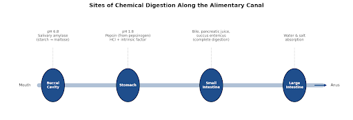
Figure 1: Sites of chemical digestion along the alimentary canal, with pH and enzyme details.
Buccal cavity: digestion begins the moment you chew. Salivary amylase breaks down roughly 30% of dietary starch into a simpler sugar, maltose, at an optimum pH of 6.8.
Stomach: gastric glands contain peptic cells, which secrete inactive pepsinogen, and parietal (oxyntic) cells, which release hydrochloric acid (HCl) and intrinsic factor. The highly acidic environment (pH 1.8) activates pepsinogen into pepsin, which splits proteins into proteoses and peptones.

Small intestine: complete chemical digestion happens here, where the food mix meets three major secretions:
- Bile: produced by the liver and stored in the gallbladder. It contains bile salts that emulsify large fat droplets into tiny micelles, but no enzymes.
- Pancreatic juice: contains inactive enzymes such as trypsinogen, chymotrypsinogen, and procarboxypeptidase. Enterokinase (from the intestinal mucosa) activates trypsinogen into trypsin, which then switches on the remaining enzymes.
- Succus entericus: the intestinal juice itself, containing final-stage enzymes — disaccharidases, dipeptidases, lipases, and nucleosidases — that finish breaking down the remaining nutrients.
Absorption Mechanisms
Once food is broken down into its simplest units, it moves across the intestinal mucosa to enter the blood or lymph, through four distinct mechanisms:
Best Courses for You

JEE

NEET

Foundation JEE

Foundation NEET

CBSE
- Simple diffusion: small amounts of glucose, amino acids, and electrolytes like chloride ions pass into the blood along their concentration gradient.
- Facilitated transport: glucose and certain amino acids cross the membrane with the help of specific carrier proteins.
- Active transport: nutrients move against their concentration gradient using metabolic energy (ATP) — the primary route for sodium ions, amino acids, and glucose.
- Fat absorption: fatty acids and glycerol are insoluble in water, so they are first packaged into water-soluble micelles, move into intestinal cells, and are re-formed into protein-coated fat globules called chylomicrons. These enter the lymph vessels (lacteals) inside the intestinal villi before joining the venous circulation.
Do Check: NEET 2026 Cut Off For BSc Nursing Government Colleges
Breathing and Exchange of Gases
Every breath has one purpose: supplying oxygen while removing carbon dioxide. This depends on pressure changes inside the chest that draw air into the lungs, where gases cross into the bloodstream.
Mechanism of Breathing
Breathing relies on a pressure gradient between atmospheric air and the air inside the thoracic cavity, driven by the diaphragm and the intercostal muscles between the ribs.
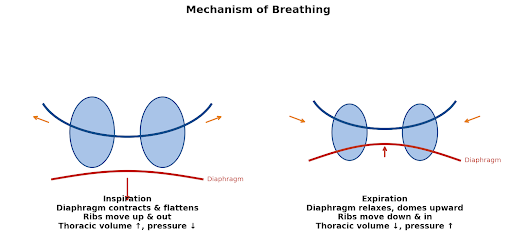
Figure 2: Inspiration expands the thoracic cavity; expiration lets it passively recoil.
Inspiration: an active process triggered by contraction of the diaphragm (which moves downward) and the external intercostal muscles (which lift the ribs up and outward). This expands thoracic volume; the resulting drop in intra-pulmonary pressure below atmospheric pressure pulls air into the lungs.
Expiration: a passive process during normal quiet breathing. The diaphragm and external intercostal muscles relax, the thoracic cavity contracts back to its original volume, intra-pulmonary pressure rises above atmospheric pressure, and air is forced out.
Respiratory Volumes and Capacities
NEET regularly tests these exact quantitative values — they are worth memorising precisely, since a capacity is always the sum of two or more volumes.
| Parameter | Definition | Average Value |
| Tidal Volume (TV) | Air inspired or expired during a normal, quiet breath | 500 mL |
| Inspiratory Reserve Volume (IRV) | Additional air a person can inspire by forceful inspiration | 2500 – 3000 mL |
| Expiratory Reserve Volume (ERV) | Additional air a person can expire by forceful expiration | 1000 – 1100 mL |
| Residual Volume (RV) | Air remaining in the lungs even after forcible expiration | 1100 – 1200 mL |
| Vital Capacity (VC) | Maximum air a person can breathe out after a forced inspiration | TV + IRV + ERV ≈ 4000 mL |
| Total Lung Capacity (TLC) | Total air the lungs hold at the end of a forced inspiration | VC + RV ≈ 5000 – 6000 mL |
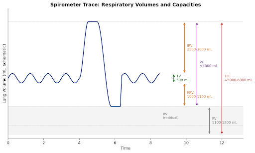
Figure 3: Spirometer trace showing how each volume and capacity is measured.
Gas Transport Mechanisms
Gases move across the ultra-thin respiratory membrane by simple diffusion, driven by partial pressure gradients (pO₂ and pCO₂).
Oxygen transport: around 97% of oxygen binds reversibly to the iron-bearing protein haemoglobin inside red blood cells, forming oxyhaemoglobin; the remaining 3% travels dissolved directly in blood plasma. Oxygen binding follows a sigmoidal curve, the oxygen-haemoglobin dissociation curve.
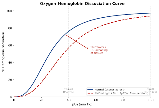
Figure 4: The dissociation curve shifts right under acidosis, high pCO₂, and elevated temperature — exactly the conditions found in active tissue.
High pO₂ in the alveoli favours oxyhaemoglobin formation, while low pO₂, high pCO₂, high H⁺ concentration (low pH), and elevated temperature in peripheral tissues trigger the release of oxygen — shifting the curve to the right.
Carbon dioxide transport: CO₂ travels through the blood in three forms:
- As bicarbonate ions (HCO₃⁻) in blood plasma: 70% (facilitated by the enzyme carbonic anhydrase).
- Bound to haemoglobin as carbaminohaemoglobin: 20–25%.
- Dissolved directly in blood plasma: 7%.
Do Check: Human Physiology
Body Fluids and Circulation
Onutrients and oxygen enter the bloodstream, the heart takes over. Every heartbeat keeps those materials moving through the body, delivering vital supplies to peripheral tissues while hauling away metabolic waste.1 Blood Composition and Groups
Blood is a specialised fluid tissue consisting of liquid plasma (55%) and formed elements (45%) — erythrocytes (RBCs), leukocytes (WBCs), and platelets (thrombocytes).
ABO system: determined by A and B antigens on RBC surfaces. Type O blood lacks both surface antigens, making it the universal donor; type AB blood lacks anti-A and anti-B antibodies in the plasma, making it the universal recipient.
Rh factor: an additional surface antigen found on the RBCs of roughly 80% of humans (Rh⁺). If an Rh⁻ mother carries an Rh⁺ fetus during a second pregnancy, her anti-Rh antibodies can cross the placenta and destroy fetal RBCs — a life-threatening condition called erythroblastosis fetalis.
Structure of the Heart and the Cardiac Cycle
The human heart is a muscular, four-chambered organ that pumps blood through synchronised electrical and mechanical phases. The cardiac cycle describes the sequence of events during a single heartbeat, lasting approximately 0.8 seconds.
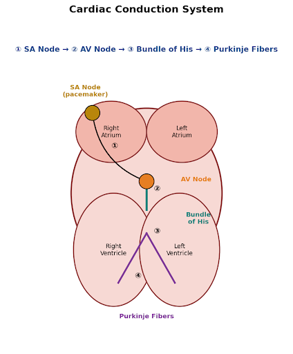
Figure 5: The cardiac conduction pathway — SA node → AV node → Bundle of His → Purkinje fibers.
- Joint diastole (0.4 s): all four chambers are relaxed. Blood flows from the vena cava and pulmonary veins into the atria, then through the open tricuspid and bicuspid valves into the ventricles.
- Atrial systole (0.1 s): the SA node fires an electrical impulse that spreads across the atria, causing them to contract and pump an additional 30% of blood into the ventricles.
- Ventricular systole (0.3 s): the electrical signal passes through the AV node and the Bundle of His to the Purkinje fibers, triggering ventricular contraction. Rising pressure closes the tricuspid and bicuspid valves, creating the first heart sound (“lub”). Soon after, the semilunar valves fly open, forcing blood into the pulmonary artery and aorta.
- Ventricular diastole: as the ventricles relax, intra-ventricular pressure drops. The semilunar valves snap shut to prevent backflow, producing the second heart sound (“dub”).
The volume of blood pumped out by each ventricle during a single contraction is the stroke volume (≈ 70 mL). Multiplying this by the heart rate gives the total cardiac output:
Cardiac Output = Stroke Volume × Heart Rate = 70 mL × 72/min ≈ 5040 mL/min ≈ 5 L/min
4. Excretory Products and Their Elimination
Every kidney contains close to a million nephrons, each working like a tiny filtration unit. Together they clean the blood continuously through three major steps to remove nitrogenous waste. Because humans are ureotelic, the liver first converts highly toxic ammonia into urea, which the kidneys then isolate and extract.
Nephron Structure and Urine Formation
Glomerular filtration: blood enters the glomerulus under high pressure via the wide afferent arteriole. Water and small solutes are forced across the three-layered filtration membrane into Bowman's capsule — a non-selective process called ultrafiltration. The average glomerular filtration rate (GFR) is 125 mL/min, around 180 litres of filtrate per day.
Tubular reabsorption: since the body cannot afford to lose 180 litres of fluid daily, nearly 99% of the filtrate is reabsorbed as it travels through the renal tubules. The Proximal Convoluted Tubule (PCT) reabsorbs nearly all essential nutrients, glucose, amino acids, and 70–80% of electrolytes and water.
Tubular secretion: cells in the renal tubules selectively secrete ions like H⁺, K⁺, and ammonia (NH₃) back into the moving filtrate, playing a vital role in maintaining the body's ionic balance and blood pH.
4The Counter-Current Mechanism
To conserve water, mammals excrete hypertonic urine — urine far more concentrated than blood plasma. This concentration process relies on a counter-current mechanism driven by two parallel, U-shaped tubes in the renal medulla: the Loop of Henle and the Vasa Recta.
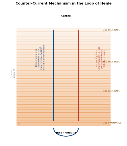
Figure 6: The counter-current mechanism builds an osmotic gradient from 300 mOsmol/L at the cortex to nearly 1200 mOsmol/L deep in the medulla.
- The descending limb is permeable to water but impermeable to electrolytes. As filtrate flows downward, water moves out into the concentrated medullary interstitium, and the filtrate becomes progressively more concentrated.
- The ascending limb is impermeable to water but allows the transport of electrolytes (NaCl) into the interstitium. As filtrate flows upward, it loses salt and becomes dilute.
The result is a steep osmotic gradient inside the renal medulla, starting at about 300 mOsmol/L near the cortex and rising to nearly 1200 mOsmol/L deeper inside the kidney. That difference allows water to leave the collecting duct, producing concentrated urine.
NCERT-Based MCQs
Q1. Which of the following cells in the gastric glands secrete intrinsic factor required for the absorption of vitamin B₁₂?
(A) Peptic cells
(B) Oxyntic cells
(C) Zymogen cells
(D) Goblet cells
Answer & Explanation: Oxyntic (parietal) cells secrete both HCl and intrinsic factor, which is essential for vitamin B₁₂ absorption in the ileum.
NCERT Reference: Class 11, Page 262
Q2. If the absolute Tidal Volume of a healthy human is 500 mL and the Expiratory Reserve Volume is 1000 mL, what is the total volume of air a person can expire forcefully after a normal, unforced inspiration?
(A) 1500 mL
(B) 1000 mL
(C) 2500 mL
(D) 3500 mL
Answer & Explanation: The total volume expired after a normal inspiration equals TV + ERV = 500 + 1000 = 1500 mL.
NCERT Reference: Class 11, Page 272
Q3. What is the correct sequence of electrical impulse conduction through the human heart?
(A) AV Node → SA Node → Purkinje Fibers → Bundle of His
(B) SA Node → AV Node → Bundle of His → Purkinje Fibers
(C) SA Node → Purkinje Fibers → AV Node → Bundle of His
(D) AV Node → Bundle of His → SA Node → Purkinje Fibers
Answer & Explanation: The electrical impulse starts at the SA node, moves to the AV node, travels down the Bundle of His, and branches out into the Purkinje fibers.
NCERT Reference: Class 11, Page 284
Q4. Which section of the nephron is completely impermeable to water molecules even under high osmotic pressure?
(A) Descending limb of Loop of Henle
(B) Proximal Convoluted Tubule
(C) Ascending limb of Loop of Henle
(D) Collecting Duct
Answer & Explanation: The ascending limb of the Loop of Henle is impermeable to water but allows active or passive transport of electrolytes.
NCERT Reference: Class 11, Page 294
Q5. A severe decrease in glomerular blood pressure triggers the Juxtaglomerular (JG) cells to release which enzyme into the bloodstream?
(A) Rennin
(B) Renin
(C) Angiotensinogen
(D) Erythropoietin
Answer & Explanation: A drop in GFR activates JG cells to release Renin (spelled with a single ‘n’), which kicks off the RAAS pathway to restore blood pressure.
NCERT Reference: Class 11, Page 297
Q6. Which constituent of blood plasma is primarily responsible for maintaining normal blood osmotic pressure?
(A) Fibrinogen
(B) Globulins
(C) Albumins
(D) Serum
Answer & Explanation: Albumins are the primary plasma proteins responsible for maintaining the colloid osmotic balance of blood.
NCERT Reference: Class 11, Page 279
Q7. What happens to the oxygen-haemoglobin dissociation curve when a patient experiences severe tissue hypoxemia combined with lactic acidosis?
(A) Shifting to the left
(B) Shifting to the right
(C) Becoming a straight linear progression
(D) Shifting completely flat
Answer & Explanation: Increased H⁺ concentration (acidosis), elevated pCO₂, and higher temperature all reduce haemoglobin's affinity for oxygen, shifting the dissociation curve to the right to favour oxygen unloading.
NCERT Reference: Class 11, Page 274
Q8. The second heart sound, “dub,” is caused by the sudden closure of which heart valves?
(A) Tricuspid valve
(B) Bicuspid valve
(C) Semilunar valves
(D) Mitral valve
Answer & Explanation: Closure of the semilunar valves at the beginning of ventricular diastole produces the second heart sound.
NCERT Reference: Class 11, Page 285
Q9. Which of the following substances is completely reabsorbed from the glomerular filtrate through active transport under normal physiological conditions?
(A) Urea
(B) Water
(C) Glucose
(D) Nitrogenous waste
Answer & Explanation: Glucose and amino acids are fully reabsorbed via active transport in the PCT, ensuring zero loss in urine under normal conditions.
NCERT Reference: Class 11, Page 293
Q10. Which enzyme converts inactive trypsinogen into active trypsin within the small intestine?
(A) Pepsin
(B) Chymotrypsin
(C) Enterokinase
(D) Steapsin
Answer & Explanation: Enterokinase, secreted by the intestinal mucosa, activates pancreatic trypsinogen into trypsin.
NCERT Reference: Class 11, Page 262
Do Check: NEET Chemistry Organic Reactions
Common Pitfalls to Avoid in NEET Biology
Watch out for these frequent mistakes when practising Human Physiology problems:
- Mixing up Renin and Rennin: Renin (single ‘n’) is a proteolytic kidney enzyme involved in blood pressure regulation. Rennin (double ‘n’) is a milk-curdling digestive enzyme found in the gastric juice of infants.
- Confusing volume and capacity formulas: a capacity is always a sum of two or more volumes (e.g., Functional Residual Capacity = ERV + RV).
- Misinterpreting heart sound triggers: heart sounds are caused by the closure of valves, never their opening. “Lub” marks closure of the AV valves; “dub” marks closure of the semilunar valves.
- Skipping diagram labels: NEET frequently adapts line diagrams directly from the textbook and replaces structural labels with letters (A, B, C, D). Practice identifying every structure on a completely blank diagram.
How Infinity Learn Helps with Human Physiology
Human Physiology includes processes that are difficult to imagine from static textbook diagrams alone. Infinity Learn explains topics such as the cardiac cycle, nephron function, and gas exchange through animated lessons, followed by chapter-wise practice questions and NCERT-based tests. You can learn a concept, solve questions on it immediately, and identify weak areas without switching between different resources.
Conclusion
Human Physiology becomes much easier once you stop treating every chapter separately. Digestion, circulation, breathing, and excretion constantly interact with one another. When you understand those connections, remembering individual facts becomes much easier during revision. Spend your study time identifying how these organ systems communicate, instead of trying to memorise biological figures in isolation.
course
No courses found
Which topics in human physiology are most important for NEET?
While all chapters are testable, the highest question density typically comes from the counter-current mechanism in Excretion, the cardiac cycle and ECG interpretations in Body Fluids and Circulation, respiratory capacities in Breathing and Exchange of Gases, and enzymatic actions within Digestion and Absorption.
How many questions come from human physiology in NEET?
On average, you can expect between 12 and 14 questions from this unit, making up roughly 15% of the entire Biology section — the highest-weightage human biology block in the syllabus.
What is the best way to learn diagrams for NEET Biology?
The most effective approach is active recall. Photocopy or trace the NCERT diagrams with the labels blanked out, and practice writing down the names of the parts from memory. Pay close attention to the arrows indicating direction of flow in circulatory and excretory diagrams.
Should I study human physiology from NCERT only?
Yes. NCERT should be your starting point and your main reference, since most NEET questions in Human Physiology come directly from it. Use reference materials only to clarify complex mechanisms — like the Loop of Henle concentration steps — never to memorise bloated, non-syllabus medical facts.
How do I remember enzyme names and functions easily?
Group the enzymes by their organ of origin and target substrate. Most digestive enzymes are named after the molecule they break down, followed by the suffix “-ase” (e.g., maltase breaks down maltose, lipases break down lipids). Keeping a running chart of active pH environments also simplifies the learning process.