
Table of Contents
A microscope is a scientific tool that is used to study items that are too small for the naked eye to see. Microscopy seems to be the science of using a microscope to study small items and structures. Unless assisted by a microscope, microscopic means invisible to naked sight. Microscopes originate in a range of shapes and sizes, and they can be defined in a range of ways. One method is to describe how an instrument interacts with a sample and produces images, such as transmitting a beam of light or electrons through a sample along its optical path, detecting photon emissions from a sample, or scanning over and a short distance from the sample surface with a probe.
A brief outline
A microscope is a device that magnifies items that would otherwise be too small to see, resulting in a picture that makes the object appear larger. The majority of cell photographs are taken with a microscope, and these images are referred to as micrographs. Compound microscopes, which have many lenses, are the more advanced tools that we commonly think of as microscopes. These lenses can bend light to generate a considerably more enlarged image than a magnifying glass because of the way they are arranged.
The arrangement of the lenses in a compound microscope having two lenses has an intriguing result: the orientation of the picture you see is inverted in regard to the actual object you’re investigating. For example, if you looked through a microscope at a piece of newsprint with the letter “e” on it, the image you would see would be “.” Because more advanced compound microscopes have an extra lens that “re-inverts” the image returns to its old condition, they may not create an inverted image.
Important concepts
Magnification refers to how much larger an object appears when viewed via a microscope (or a pair of lenses). Light microscopes, for example, commonly used in high schools and colleges magnify objects up to 400 times their true size.
The lowest distance by which two can be split and still be recognized as independent objects is the resolution of a microscope or lens. The lower this value, the stronger the resolving power and the better the image clarity and detail.
The Basic Microscope Principle
When a material is placed within the focus of a basic microscope, a virtual, erect, and enlarged image is obtained at the shortest possible distance of distinct vision from the eye that is placed at the lens.
Applications
- It’s prevalent among watchmakers since it allows them to see a magnified image of the tiniest parts.
- Jewelers use it to get a magnified image of the fine elements of the jewelry.
- In most educational institutions’ laboratories, such as schools and colleges, a basic microscope is used.
- Used by dermatologists (skin specialists) to diagnose various skin conditions.
Compound Microscope Principle
The compound microscope works on the idea that combining lenses increases the magnification of the sample. The material is initially observed in the tube as a primary picture, then in the eyepiece.
Applications
- To study bacteria and viruses.
- In forensic laboratories, It is used.
- It’s also employed in the field of metallurgy.
The term “compound microscopes” refers with many lenses. It has two optical parts: an objective lens and an eyepiece or ocular lens, which are made up of a combination of lenses.
Electron Microscope
An electron microscope is used as an accelerated electron beam as its source of illumination. Because the images can be amplified in nanometers, it is a particular sort of microscope with excellent image resolution.
Electron microscopes are divided into two categories:
- The transmission electron microscopes (TEM)
- SEM stands for scanning electron microscopes (SEM)
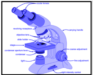
The pieces of a basic microscope are listed below, along with their functions:
- The ocular is the lens at the top of the microscope that is used to examine the samples. The magnification ranges from 10X to 15X.
- It is supported by the base.
- The eyepiece is connected to the objective lenses by a tube.
- Colour-coded objective lenses with magnifications of 10X, 40X, and 100X are available. The shortest lenses are those with the lowest power, and the longest lenses are those with the highest power.
- Nosepiece that rotates: The turret is another name for this structure. It may be rotated while examining the samples and is used to hold various objective lenses.
- The diaphragm is used to regulate the quantity of light that enters the stage.
- The stage is the platform on which the slides with specimens are placed.
- Stage clips are used to keep the slides in position on the stage.
- It’s utilized to focus on scanning using the coarse adjustment knob.
- It’s utilized to focus on oil with the fine adjustment knob.
- The arm is used to sustain the tube and attaches to the microscope’s base.
- The main power switch is used to turn the microscope on and off.
- Condenser: A condenser is a device that focuses light on a sample using 400X power lenses.
FAQs:
What's the difference between a low-powered microscope and a high-powered microscope?
The primary distinction between low-powered and high-powered microscopes is that a high-powered microscope is utilized for resolving smaller features due to the high magnification of the objective lenses. Low-powered objectives, on the other hand, have the greatest depth of focus. The depth of focus decreases as the power is increased.
In a microscope, what is the depth of focus?
The distance between the objective lens as well as the sample plane in a microscope is known as the depth of focus. The quality of attention affects the depth of focus, which differs from person to person.
In a microscope, and what's the depth of field?
In a microscope, the depth of field is defined as the distance between the closest object plane in focus and the farthest plane in the same focus. The depth of field in microscopes is extremely small, measured in microns.
In a microscope, what is the field of view?
The diameter of the lit circle seen through the eyepiece defines the field of view in a microscope. The arena of view shrinks as the magnification rises.
