Table of Contents
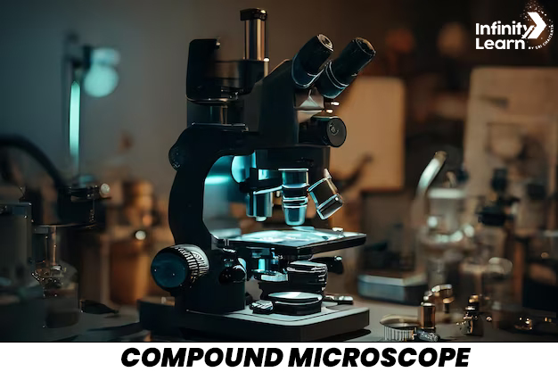
Have you ever wondered how scientists and researchers can see tiny things like cells and bacteria? The answer is the compound microscope! This amazing tool allows us to magnify small objects and see them in great detail.
In this article, we’ll explore the compound microscope, its parts, and how it works. We’ll also learn about the uses of compound microscope and how it has helped us understand the world around us.
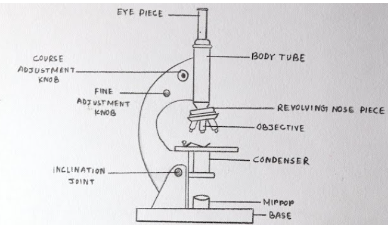
Let’s start with a compound microscope diagram to get a better idea of its structure. The microscope has two main lenses: the objective lens and the eyepiece lens. The objective lens is located near the object you want to observe, while the eyepiece lens is where you look through.
By understanding the parts of compound microscope and how they work together, you’ll be able to use this tool effectively and make amazing discoveries!

Compound Microscope Ray Diagrams
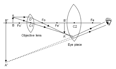
Compound Microscope consists of the eyepiece positioned for direct viewing, which further magnifies the image produced by the objective lenses near the specimen under observation. Objective lenses with various magnification levels, such as 4x, 10x, 40x, and 100x, contribute to a comprehensive examination of the specimen, achieving total magnifications exceeding 1000 times the actual size.
Below given is the compound microscope ray diagram:

The stage provides a platform for securing the specimen during observation, often equipped with clips for stability. Coarse and fine adjustment knobs ensure precise focusing on the specimen, enabling a clear and detailed view. This precision in focusing is essential for revealing the fine structures of cells, microorganisms, and other microscopic entities.
Below the stage, a condenser and illuminator are pivotal components. The condenser focuses and directs light onto the specimen, enhancing visibility. The illuminator, whether integrated into the microscope or an external light source, ensures proper lighting, making it easier to observe the tiny object.
The compound microscope ray diagram illustrates how light travels through the lenses to magnify the specimen. The objective lens forms a real, inverted, and magnified image of the object, which is then further magnified by the eyepiece lens. This combination of lenses allows for significant magnification, enabling us to see the intricate details of microscopic structures.
In widespread use across fields like biology, medicine, and materials science, compound microscopes are indispensable tools for scientific exploration and education. Their versatility and ability to resolve fine details empower researchers to delve into the hidden world of cells, microorganisms, tissues, and other microscopic structures, fostering a deeper understanding of the microscopic realm.
Parts of Compound Microscope
Optical Parts
- Eyepiece (Ocular Lens): The eyepiece is similar to a magnifying glass that makes things appear larger when you look through it.
- Objective Lenses: These special lenses zoom in on tiny objects, allowing for a clearer and more detailed view.
- Condenser: Think of the condenser as a flashlight that illuminates the small objects, making them easier to see.
Non-Optical Parts
- Stage: The stage is like a small table where you place the objects you want to observe.
- Coarse and Fine Adjustment Knobs: These knobs help in focusing on the tiny objects. The coarse knob moves things quickly, while the fine knob allows for precise focusing.
- Illuminator: Similar to a lamp, the illuminator is the light source that aids in better visibility of the tiny objects.
- Diaphragm: Acting like a curtain, the diaphragm controls the amount of light reaching the objects, improving visibility.
- Base: The base serves as the foundation, keeping the microscope stable and secure.
Working Principle of Compound Microscope
A compound microscope is a remarkable tool that employs multiple lenses to magnify small objects, unveiling the secrets of the microscopic world. Scientists, researchers, and students widely use this ingenious device to explore the intricate details of tiny things that are invisible to the naked eye.
Check the compound microscope diagram below:

At the core of the compound microscope are its lenses—the objective lenses and the eyepiece. The objective lenses, positioned close to the tiny object being observed, come in different magnification powers, such as 4x, 10x, 40x, and 100x. The eyepiece, placed close to your eye, further magnifies the image produced by the objective lenses.
The tiny object to be observed is carefully placed on a flat stage, equipped with clips to keep it steady. Using adjustment knobs, scientists can focus on the object, bringing it into clear view. Additionally, a built-in or external light source illuminates the object, making it easier to see the details.
The chosen objective lens captures a magnified image of the tiny object, and then the eyepiece further magnifies that image. The result is a significantly enlarged and detailed image that you can observe by looking through the eyepiece.
This compound microscope, with its ability to magnify and reveal tiny details, is like a superhero in the world of science. It allows us to explore the hidden wonders of cells, microorganisms, and other small structures, contributing to our understanding of the microscopic realm and opening new avenues for scientific discovery.
Advantages and Disadvantages of Compound Microscope
Advantages of Compound Microscopes
- Magnification Power: Compound microscopes can magnify tiny objects, allowing scientists to observe intricate details.
- Clarity in Observations: They provide clear and detailed images, aiding researchers in the thorough examination of specimens.
- Versatility in Applications: These microscopes are suitable for various tasks, including studying living organisms, medical research, and analysing materials.
- Enhanced Depth Perception: They enable observers to perceive objects in a three-dimensional manner, adding depth to their observations.
- Adjustable Lighting: Users have the flexibility to control the amount of light, optimising visibility during observations.
Disadvantages of Compound Microscopes
- Learning Curve: Operating compound microscopes may require some practice and familiarity, making them challenging for beginners.
- Limited Portability: Due to their size, these microscopes are not easily portable, limiting their use to specific locations.
- Maintenance Requirements: Regular care is essential to keep the microscope in good working condition, ensuring cleanliness of lenses and components.
- Potential Cost Concerns: High-quality compound microscopes can be expensive, posing financial challenges for individuals or institutions with limited budgets.
- Reduced Field of View: At higher magnifications, the visible area may become limited, making it difficult to study larger specimens comprehensively.
FAQs on Compound Microscope
What are two types of Compound Microscope?
There are many types of Compound Microscope. These include biological microscopes, polarising microscopes, phase contrast microscopes, and fluorescence microscopes. Each of them has a different function.
Why is a Compound Microscope important?
The compound microscope is vital in unveiling the microscopic world. It enriches life by revealing otherwise unseen wonders and expanding knowledge
What are some common applications of compound microscopes?
Common applications include biological research, medical diagnostics, educational purposes, quality control in industries, and forensic analysis.
Who invented compound microscope?
The invention of the microscope opened up new ways for scientists to learn about the body and diseases. Although it's uncertain who exactly created the first microscope, Zacharias Janssen, a Dutch maker of eyeglasses born in 1585, is recognized for crafting one of the earliest compound microscopes (which used two lenses) around 1600.


