Table of Contents
Important Topic of Biology: Cell Cycle
Definition
The cell cycle is a series of ordered events that involves the growth of the cell and cell division which produces two new daughter cells. All cells reproduce by dividing into two more new daughter cells from each parent cell.
A brief note
Cell division
Cell division is a very important process in all living organisms and this repeating cell division process is called the cell cycle. During the cell division, growth of the cell and DNA replication takes place. This ordered event of dividing the cells and forming two new cells is called a cell cycle.
The cell cycle is divided into two phases which are:
- Interphase
- Mitosis phase or M phase
Interphase: The interphase is also called the resting phase, is the time during which the cells are preparing for cell division by undergoing cell growth and replication of the DNA in a proper sequence. The interphase is divided further into three other phases:
- G1 Phase (Gap 1)
- S Phase (synthesis)
- G2 phase (gap 2)
G1 phase :
During the G1 phase, the cells are metabolically active and continuously grow and increase in size but do not replicate the DNA. G1 phase takes place between mitosis and DNA replication.
S Phase (Synthesis):
This is a phase or period where replication or synthesis of DNA occurs. During the Synthesis period, the amount of DNA per cell will double in this phase.
G2 phase:
During this phase, there is a gap between mitosis and DNA synthesis, the cell will continue to grow and proteins are synthesized to make sure that everything is ready to enter the mitosis stage.
There are some cells in the organisms that do not seem to exhibit cell division because of injury or cell death. The cells which do not divide exit the G1 phase to enter an inactive stage called the Quiescent stage which is also called the G0 phase of the cell cycle.
M Phase: It is the phase where the cell growth stops and the cell energy will be distributed into two new daughter cells. After cell division, each new daughter cell will start the interphase of the new cycle making another cell cycle.
The mitosis will be divided into four stages of nuclear division, also called karyokinesis. The karyokinesis is further divided into four stages, which are:
- Prophase
- Metaphase
- Anaphase
- Telophase
Let us know more about each stage of karyokinesis:
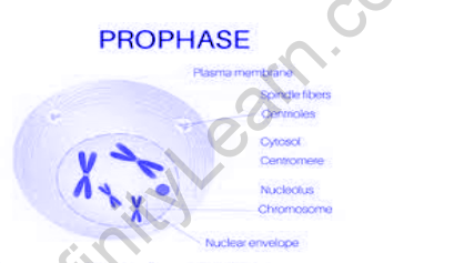
Prophase
The first stage of karyokinesis or mitosis is called prophase. It is a process that will separate the duplicated genetic material which is present in the parent cell into two daughter cells that are identical. The new DNA molecules formed are intertwined. The prophase is that whenever chromosomal material commences condensing. During the chromatin condensation process, the chromosomal material becomes untangled. The centrosome, which has been reproduced during the S phase of interphase, now begins to move towards the cell’s opposite poles. Prometaphase, the second phase of mitosis, follows prophase.
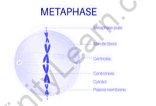
Metaphase
The second phase of mitosis begins when the nuclear envelope completely disintegrates, and the chromosomes are spread throughout the cell’s cytoplasm. Chromosome condensation is complete at this point, and they can be seen clearly under the microscope. This is the stage at which the morphology of chromosomes can be studied the most easily. The metaphase chromosome is made up of two sister chromatids held together by the centromere at this stage. Kinetochores are small disc-shaped structures on the surface of centromeres. The spindle fibers are attached to the chromosomes that are moved into position at the cell’s center by these structures. As a result, all of the chromosomes come to lie at the equator during metaphase, with one chromatid of each chromosome connected to spindle fibers from one pole and its sister chromatid connected to spindle fibers from the opposite pole by its kinetochore.
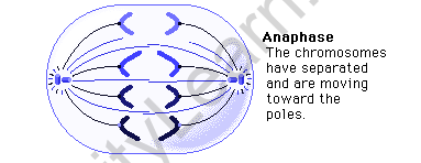
Anaphase
At the beginning of anaphase, each chromosome on the metaphase plate is split into two daughter chromosomes. Chromatids, furthermore identified as daughter chromosomes, are also the daughter chromosomes of a cell. The future daughter nuclei start migrating towards the mother nucleus. The two polar opposites each chromosome moves away from the others. Each chromosome’s centromere is positioned on the equatorial plate. remains primarily directed at the pole and, as a result, at the front edge, with the chromosome arms trailing behind.
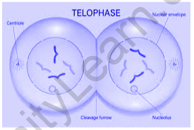
Telophase
The chromosomes that have reached their respective poles decondense and lose their individuality at the beginning of the final stage of karyokinesis, telophase. Each set of chromatin material tends to collect at each of the two poles, and individual chromosomes can no longer be seen. As chromosomes reach the cell poles, a nuclear envelope forms around each set of chromatids, nucleoli reappear, and chromosomes begin to decondense back into the expanded chromatin of interphase.
Cytokinesis
Mitosis typically involves not only the segregation of duplicated chromosomes into daughter nuclei (karyokinesis), but also the division of the cell into two daughter cells (cytokinesis), at which point cell division is complete. The appearance of a furrow in the plasma membrane in an animal cell accomplishes this. The furrow deepens over time and eventually joins in the midpoint, dividing the cell cytoplasm in half. Plant cells, on the other contrary, have a relatively inextensible cell wall, so they go through cytokinesis in a different way. Wall formation in plant cells begins in the cell’s center and spreads outward to meet the cell’s existing lateral walls. The formation of a simple precursor known as the cell plate is the first step in the formation of the new cell wall, which is the middle lamella between the walls of two adjacent cells.
Meiosis
The fusion of two gametes, each with a complete haploid set of chromosomes, is considered necessary for sexual reproduction to produce offspring. Diploid cells with specialized functions produce gametes. The production of haploid daughter cells results from a specialized type of cell division that reduces the chromosome number by half. Meiosis is the specific type of division. In the life cycle of sexually reproducing organisms, meiosis ensures the production of the haploid phase, whereas fertilization restores the diploid phase. Meiosis is a process that takes place during gametogenesis across both plants and animals.
Meiosis comprises two completely separate nuclear and cell division cycles called meiosis I and meiosis II.
Meiosis I
Prophase I:
When compared to mitosis, the prophase of the first meiotic division is typically longer and more complex. It has been further subdivided into the following five phases based on chromosomal behavior: Leptotene, Zygotene, Pachytene, Diplotene, and Diakinesis.
Metaphase I:
On the equatorial plate, the bivalent chromosomes align. Microtubules from complete opposites of the spindle attach to homologous chromosomes’ kinetochore.
Anaphase I:
Sister chromatids remain attached at their centromeres as homologous chromosomes separate.
Telophase I:
The nuclear membrane and nucleolus reappear, followed by cytokinesis and the formation of a dyad of cells. Regardless of the fact that chromosomes disperse in many cases, they do not reach the interphase nucleus’s extremely extended state. Interkinesis is a stage that occurs between the two meiotic divisions and lasts only a few minutes.
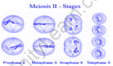
Meiosis II
Prophase II:
Meiosis II begins soon after cytokinesis, usually before the chromosomes have fully extended. Meiosis II, in contrast to meiosis I, resembles a normal mitosis. By the end of prophase II, the nuclear membrane has disappeared completely. The chromosomes are compacted once more.
Metaphase II:
The chromosomes align at the equator at this juncture, and microtubules from opposite poles of the spindle attach to the sister chromatids’ kinetochores.
Anaphase II:
It tends to start with the centromeres of each chromosome splitting around the same time, allowing them to move toward opposite poles of the cell by shortening microtubules attached to kinetochores.
Telophase II:
Meiosis finally ends with telophase II, when the two groups of chromosomes are once again wrapped in a nuclear envelope; cytokinesis then takes place, As a result, a tetrad of cells, or four haploid daughter cells, is formed.
Also read: Important Topic Of Biology: Microbodies
FAQs
Why do cells need to divide?
It is absolutely essential for cells to divide in order for you to grow and for your cuts to heal. It’s also extremely important for cells to stop dividing whenever they’re supposed to. Cancer develops when a cell fails to prevent dividing when it is supposed to. Skin cells, for example, are constantly dividing.
How many times can a cell go through the cell cycle?
The Hayflick Limit is a concept that aids in the comprehension of cellular aging mechanisms. According to the theory, a normal human cell can only replicate and divide forty to sixty times before it can no longer divide and dies from apoptosis, or programmed cell death.
How fast do cells replicate?
The S phase usually takes 5 to 6 hours for cells to complete. G2 is the shortest of the three phases, lasting only 3 to 4 hours in most cells. In general, the interphase period lasts between 18 and 20 hours. Mitosis, the process by which a cell prepares for and completes cell division, takes only about 2 hours.
What is the longest phase of the cell cycle?
The longest phase of the cell cycle is the interphase. The cell goes through normal growth processes while preparing for cell division during interphase. It is the longest phase of the cell cycle, with the cell spending roughly 90% of its time here.
For more visit Cell Cycle and Division – Interphase, Regulation and and Important FAQs


