Table of Contents
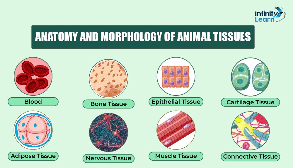
Anatomy And Morphology Of Animal Tissues: The study of anatomy and morphology of animal tissues focuses on their structure and form. Animal tissues are mainly divided into four categories: epithelial, connective, muscle, and nervous tissues. Epithelial tissues cover and protect the surfaces of organs and other body structures.
Connective tissues support and connect different parts of the body. Muscle tissues enable movement and include three types: skeletal, cardiac, and smooth muscles. Nervous tissues send signals throughout the body, allowing communication between various body parts. Each tissue type consists of specialized cells that carry out specific functions, contributing to the body’s overall organization and efficiency.

Types of Animal Tissue
During development, each cell in an organism specializes to perform a specific function. Cells with similar functions group together to form tissues. Animal tissues are broadly classified into two main types:
- Simple Tissue: A collection of cells that share the same origin and structure, working together to perform a specific function.
- Compound Tissue: A group of cells with varying structures and functions that collaborate to achieve a single purpose.
Based on shape, function, and location, tissues are categorized into four primary types:
Epithelial Tissue
Epithelial Tissue consists of sheets of cells covering body surfaces and lining internal organs. They serve to protect, absorb, transport, and excrete substances. These tissues typically lack blood vessels and are found in various shapes:
- Squamous Epithelium: Flat, thin cells that form protective layers. Examples include the lining of the mouth, lungs, and skin.
- Stratified Epithelium: Multiple layers of cells that protect against damage. Found in areas like the skin and mouth.
- Cuboidal Epithelium: Cube-shaped cells with round nuclei. Located in glands such as the pancreas and thyroid, aiding in absorption and secretion.
- Columnar Epithelium: Tall, column-like cells. Found in the intestines and other organs, helping with absorption and secretion.
Connective Tissue
Connective tissue supports and connects different tissues and organs. It consists of cells, fibers, and a matrix. Major types include:
- Adipose Tissue: Composed of large cells storing fat.
- Blood: A fluid tissue with cells suspended in plasma, essential for transporting oxygen, nutrients, and waste.
- Bones: Provide support and protection with a hard structure.
- Ligaments: Connect bones and are flexible, aiding in movement.
- Cartilage: Flexible and semi-rigid, found in areas like the ears and nose, providing support and cushioning.
Muscle Tissue
Muscle tissue are responsible for movement, consisting of long, cylindrical cells. Types include:
- Skeletal or Striated Muscles: Voluntary muscles attached to bones, characterized by a striped appearance. Found in the arms and legs.
- Smooth or Non-Striated Muscles: Involuntary muscles found in the walls of internal organs, lacking stripes.
- Cardiac Muscles: Unique to the heart, with branching cells that help pump blood rhythmically.
Nervous Tissue
Nervous tissue composed of neurons that transmit signals throughout the body. Neurons have a cell body, axon, and dendrites, enabling them to send and receive signals.
These classifications help to understand the diverse functions and structures of tissues in the body.
Structure and Functions of Animal Tissue
Below are the different types and their subtypes of animal tissues, explaining the structure and Function of each type of animal tissue:
Epithelial Tissue
This tissue covers and protects body surfaces, lines internal organs and cavities, and forms glands for secretion.
Types
- Simple Squamous: Single layer of flat cells, suited for rapid diffusion and filtration.
- Simple Cuboidal: Single layer of cube-shaped cells, involved in secretion and absorption.
- Simple Columnar: Single layer of tall, column-shaped cells, often ciliated to aid in moving substances.
- Stratified Squamous: Multiple layers of flat cells, offering protection against abrasion and water loss.
- Stratified Cuboidal: Multiple layers of cube-shaped cells, found in sweat glands and salivary glands.
- Stratified Columnar: Multiple layers of tall, column-shaped cells, relatively rare, found in the urethra and large gland ducts.
- Transitional Epithelium: Multilayered epithelium that can stretch and change shape, found in the urinary bladder.
Connective Tissue
This tissue supports, protects, and binds other tissues, providing structure and function to the body.
Types
- Loose Connective Tissue: Features a loose arrangement of fibers and cells, providing support and flexibility.
- Dense Regular Connective Tissue: Contains tightly packed collagen fibers in parallel bundles, offering strength and resistance to tension.
- Dense Irregular Connective Tissue: Contains tightly packed collagen fibers arranged randomly, providing strength and resistance to tension in multiple directions.
- Elastic Connective Tissue: Has a high proportion of elastic fibers, allowing for stretching and recoil.
- Cartilage: A specialized connective tissue with a firm matrix, offering support and flexibility.
- Bone: A specialized connective tissue with a hard matrix, providing support and protection.
- Blood: A fluid connective tissue composed of cells and plasma, essential for transporting oxygen, nutrients, and waste products.
Muscular Tissue
This tissue enables movement, maintains posture, and generates heat.
Types
- Skeletal Muscle: Striated muscle fibers arranged in bundles, controlled voluntarily.
- Cardiac Muscle: Striated muscle fibers arranged in a network, controlled involuntarily.
- Smooth Muscle: Non-striated muscle fibers arranged in sheets, controlled involuntarily.
Nervous Tissue
This tissue coordinates and controls body functions, including sensation, thought, and movement.
Types
- Neurons: Specialized cells that transmit electrical signals.
- Neuroglia: Supporting cells that provide nutrients, insulation, and protection to neurons.
Difference Between Anatomy And Morphology Of Animal Tissues
The table below includes the key differences between anatomy and morphology of animal tissues:
| Difference Between Anatomy And Morphology Of Animal Tissues | ||
| Aspect | Anatomy of Animal Tissue | Morphology of Animal Tissue |
| Definition | Study of tissue structure and organization. | Study of form, shape, and development of tissues. |
| Focus | Structural relationships and organization. | Form, shape, and variations. |
| Scope | Includes both macroscopic and microscopic levels. | Primarily microscopic, but can include macroscopic aspects. |
| Objective | Understand functional and spatial relationships. | Describe and classify structural features and variations. |
| Techniques | Dissection, imaging (MRI, CT). | Microscopy, histological staining. |
| Application | Clinical diagnostics and organ function. | Developmental and evolutionary studies. |
| Examples | Liver structure, muscle tissue roles. | Tissue changes during development, cross-species comparisons. |
| Discipline | Integrates with physiology and pathology. | Integrates with developmental and evolutionary biology. |
FAQs on Anatomy And Morphology Of Animal Tissues
What is the difference between anatomy and morphology in animals?
Anatomy studies the internal structure of animals, while morphology focuses on their external form and structure.
What is animal tissue?
Animal tissue is a collection of similar cells working together to perform specific functions within the body.
What is the morphology of an animal?
The morphology of an animal refers to its overall shape, structure, and external form.
What is the anatomy and morphology of animal tissues?
Anatomy of animal tissues examines their internal organization, while morphology describes their external appearance and arrangement.


