Table of Contents
The human eye allows us to perceive the world around us through the sense of sight. The eyeball, measuring about 2.5 cm (1 in.) in diameter, is responsible for capturing and focusing light onto the retina, initiating the process of vision. To unravel the complexities of the adult eyeball, we must explore its structure, its three layers, and the remarkable processes that enable us to see the world in all its splendor.
The Three Layers of the Eyeball


The wall of the eyeball consists of three layers: the fibrous tunic, the vascular tunic, and the retina. Each layer performs crucial functions to maintain the eye’s integrity and facilitate its visual functions.
Fibrous Tunic
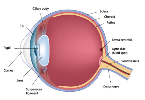
The fibrous tunic is the outermost layer of the eyeball and is composed of two parts—the anterior cornea and the posterior sclera. The cornea is a transparent coat covering the colored iris. It plays a vital role in focusing light onto the retina due to its curved shape. The sclera, often referred to as the “white” of the eye, is a dense connective tissue layer that covers the entire eyeball, except the cornea. The sclera provides shape, rigidity, and protection to the eye, and it also serves as an attachment site for the extrinsic eye muscles.


Vascular Tunic (Uvea)
The vascular tunic, also known as the uvea, lies in the middle layer of the eyeball and consists of three parts—the choroid, ciliary body, and iris. The choroid is the highly vascularised posterior portion of the vascular tunic that lines most of the internal surface of the sclera. It nourishes the posterior surface of the retina and contains melanocytes, which produce the pigment melanin, helping to absorb stray light and prevent light scattering. The ciliary body is located in the anterior portion of the vascular tunic and consists of ciliary processes and ciliary muscle. The ciliary processes secrete aqueous humor, a transparent watery fluid that nourishes the lens and cornea. The ciliary muscle is a circular band of smooth muscle that alters the shape of the lens, allowing it to adapt for near or far vision. The iris, the colored portion of the eyeball, regulates the amount of light entering the eye through the pupil. It contains circular and radial smooth muscle fibers and varies in color due to the amount of melanin present, determining eye color.
Retina (Inner tunic)
The retina, the innermost layer of the eyeball, lines the posterior three-quarters of the eye and serves as the starting point of the visual pathway. It contains specialized cells called photoreceptors—rods, and cones—which initiate the conversion of light into nerve impulses. The retina has three layers of retinal neurons—the photoreceptor cell layer, bipolar cell layer, and ganglion cell layer—separated by synaptic layers where synaptic contacts are made. The fovea centralis, a small depression in the macula lutea, is the area of highest visual acuity, responsible for sharp vision. Images focused on the retina are inverted and undergo right-to-left reversal, but our brain processes this information to perceive the world correctly. The blind spot is the region where the optic nerve exits. Image is not formed in this region as it contains no rods or cones.
Aqueous humor and Vitreous humor
Aqueous humor is the watery fluid that fills the anterior cavity (between the cornea and the iris) of the eye. It is similar to CSF (cerebrospinal fluid) in composition. The vitreous body is a soft, jellylike substance that fills the posterior chamber (between the lens and the retina) of the eyeball.


Image formation
The process of vision is a complex interplay of light and biology within the human eye. One crucial element in this intricate system is photopigments, specialized molecules that respond to light and play a pivotal role in converting visual stimuli into nerve signals.
The photopigment cycle
Photopigments are made up of two main components: the protein opsin and a derivative of vitamin A known as retinal. This cycle describes the phototransduction process that occurs when light strikes these photopigments within the retina, initiating the transformation of light into electrical signals sent to the brain.
Isomerization
In the darkness, the retinal adopts a bent shape called cis-retinal, which fits into the opsin protein. When a photon of light is absorbed by the photopigment, cis-retinal undergoes a transformation into a straightened shape called trans-retinal. This conversion, known as isomerization, is the initial step in visual transduction. As a result of this transformation, chemical changes take place in the outer segment of the photoreceptor cell.
Bleaching
After isomerization, the trans-retinal molecule separates entirely from the opsin protein in approximately a minute. This separation causes the opsin to appear colorless. Since retina is responsible for the color of the photopigment, this part of the cycle is termed “bleaching.” As the photopigment bleaches, it becomes less sensitive to light.
Conversion
Following the bleaching phase, the separated trans-retinal molecule is converted back to its original cis-retinal shape through the action of an enzyme called retinal isomerase. This conversion step prepares the photopigment for regeneration and the restoration of its sensitivity to light.
Regeneration
With cis-retinal now in its active form, it rebinds with the opsin protein, forming a functional photopigment once more. This phase of the cycle, called regeneration, is crucial as it allows the photopigment to regain its light sensitivity, enabling the eye to respond to visual stimuli effectively.
Light and dark adaptation
The eye exhibits light and dark adaptation to adjust to changing light levels. Light adaptation occurs when moving from dark surroundings to bright environments, and the eye decreases its sensitivity to light. In contrast, dark adaptation happens when moving from bright to dimly lit surroundings, and the eye increases its sensitivity to light over time. The processes of bleaching and regeneration of photopigments in the rods and cones account for these sensitivity changes during adaptation.
Summary
The human eye is a complex and remarkable organ responsible for capturing and focusing light onto the retina, enabling vision. It consists of three layers: the fibrous tunic (cornea and sclera), the vascular tunic (choroid, ciliary body, and iris), and the retina. The retina contains photoreceptors that initiate the conversion of light into nerve impulses. Image formation in the eye is akin to a camera, with the cornea and lens refracting light to focus on the retina. Accommodation, the adjustment of lens curvature by the ciliary muscle, allows us to focus on near and distant objects. The pupil’s constriction or dilation helps adapt to varying light conditions. The eye exhibits light and dark adaptation to adjust to different light levels, mediated by the bleaching and regeneration of photopigments in the rods and cones.
FAQs on Eye
How does the human eye enable us to see the world around us?
The human eye captures and focuses light onto the retina, initiating the process of vision. The retina contains specialized cells called photoreceptors (rods and cones) that convert light into nerve impulses. These impulses are then transmitted to the brain via the optic nerve, allowing us to perceive the world through the sense of sight.
What are the three layers of the eyeball and their functions?
The three layers of the eyeball are the fibrous tunic, vascular tunic (uvea), and retina. The fibrous tunic provides shape and protection to the eye, and the vascular tunic nourishes the retina and controls the amount of light entering the eye. The retina contains photoreceptors and initiates the conversion of light into nerve impulses.
What is the role of the photopigment cycle in vision?
The photopigment cycle is a crucial process that occurs within photoreceptors when light strikes them. It involves the transformation of the photopigment from cis-retinal to trans-retinal, leading to changes in the photoreceptor cell and the initiation of visual transduction. The subsequent regeneration of cis-retinal allows the photoreceptor to regain its light sensitivity.
How does the eye adapt to changes in light levels?
The eye exhibits light and dark adaptation to adjust to varying light conditions. In light adaptation, the eye decreases its sensitivity to light when moving from darkness to brightness. In dark adaptation, the eye increases its sensitivity to light when moving from brightness to dimly lit surroundings. The bleaching and regeneration of photopigments in the rods and cones account for these sensitivity changes during adaptation.
What is the fovea centralis, and why is it important for vision?
The fovea centralis is a small depression in the macula lutea, located at the center of the retina. It is responsible for the highest visual acuity and sharp vision. The fovea contains a high concentration of cones, which are responsible for color perception and detailed vision.
How does the eye focus on near and distant objects?
The process of accommodation involves the ciliary muscle altering the curvature of the lens. This adjustment allows the eye to focus on both near and distant objects by changing the lens's shape, thus refracting light appropriately onto the retina.
What are the main components of a photopigment, and what is their role in vision?
Photopigments consist of two main components: the protein opsin and the retinal molecule derived from vitamin A. When light strikes the photopigment, retinal undergoes a transformation called isomerization, initiating the process of visual transduction. The separation and subsequent regeneration of retinal and opsin play a pivotal role in the photopigment cycle.
How does the brain process the inverted images focused on the retina to perceive the world correctly?
Although images focused on the retina are initially inverted and undergo right-to-left reversal, the brain processes this information and corrects it to provide us with an upright and coherent visual perception of the world.
What is the significance of melanin in the choroid of the eye?
Melanin, produced by melanocytes in the choroid, helps absorb stray light and prevent light scattering. This process optimizes visual clarity and improves the quality of the image projected onto the retina.
What role does the iris play in regulating the amount of light entering the eye?
The iris, composed of circular and radial smooth muscle fibers, acts as a diaphragm to control the size of the pupil. It regulates the amount of light entering the eye, adapting the eye to different lighting conditions. The iris's color is determined by the amount of melanin present, which is responsible for a person's eye color.