Table of Contents
Animal Cell Diagram Class 9 Biology: The animal cell is the basic structural and functional unit of animals, serving as the building block of life for all creatures in the animal kingdom. In class 10 and 12 exams, understanding the animal cell diagram is crucial for scoring high marks. Here’s a labeled diagram along with its explanation to help you secure those marks.
Animal Cell – Introduction


The animal cell is like a tiny city inside our bodies. It has different parts, like the nucleus, which acts as the control center, and the mitochondria, which are like powerhouses producing energy. The endoplasmic reticulum and Golgi apparatus work together to make and transport proteins, while lysosomes help in cleaning up waste. Cytoplasm is where all the action happens, like a bustling marketplace where various activities take place. Knowing these organelles and their functions not only helps in acing exams but also enhances comprehension of cellular functions in living organisms. So, study this diagram well to boost your exam performance and excel in your studies.
Animal Cell Diagram Class 9 Biology
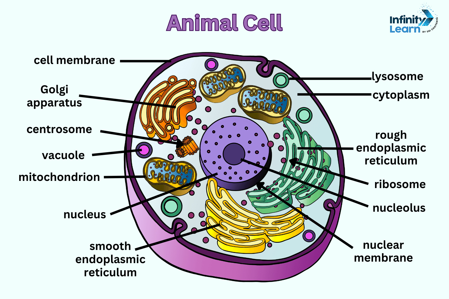


Class 9 Animal Cell Diagram: Comprehensive Guide to Structure and Functions
The animal cell, a fundamental unit of life, is a complex structure with various components. Enclosed by a cell membrane, it contains cytoplasm, where organelles like the nucleus, mitochondria, and ribosomes reside. The nucleus holds genetic material and regulates cell activities. Mitochondria produce energy, while ribosomes synthesize proteins. Other organelles like the endoplasmic reticulum, Golgi apparatus, and vesicles aid in protein synthesis, modification, and transport. Understanding the structure of an animal cell is crucial for comprehending basic biology concepts.
Also Check: CBSE Notes Class 9
Functions of Animal Cell Diagram Class 9
- Cell Membrane: A double-layered membrane primarily composed of phospholipids, enveloping the cell. It regulates the passage of substances in and out of the cell, maintaining selective permeability.
- Cytosol: The aqueous solution constituting the interior of the cell, comprised of water, ions like potassium, various proteins, and small molecules. It serves as the medium for cellular processes.
- Cytoskeleton: A network of tubules and filaments supporting cell structure and enabling movement and signaling within the cytoplasm.
- Nucleus: The central organelle housing the cell’s genetic material, DNA, situated within the nucleolus. Enclosed by the nuclear membrane, it governs cellular growth, replication, and gene expression.
- Ribosomes: Small organelles either free-floating in the cytoplasm or bound to the endoplasmic reticulum. They are instrumental in protein synthesis, translating genetic information into functional proteins.
- Endoplasmic Reticulum (ER): A complex network of membranous sacs known as cisternae, branching off from the nuclear envelope. Comprising rough and smooth types, it aids in protein synthesis, folding, and transport.
- Vesicles: Small membranous sacs involved in the transport of molecules within the cell, shuttling substances between organelles or to and from the cell membrane.
- Golgi Apparatus: The Golgi apparatus functions as a cellular “post office.” It receives proteins from the ER, modifies them, and packages them into vesicles for distribution within the cell or secretion.
- Mitochondria: Mitochondria, often called the “powerhouse of the cell,” generate ATP molecules through the oxidation of glucose and other metabolites during cellular respiration. This process provides energy for various cellular activities.
Don’t Miss
- Most Easy and Scoring Chapters of CBSE Class 9 Science
- How To Score Full Marks In CBSE Class 9 Science Exam
Benefits of Animal Cell Diagrams for Class 9
- Visual Aid: Animal cell diagrams offer a clear visual representation, aiding better understanding for Class 9 students.
- Identification: Diagrams for Class 9 help students identify different cell organelles easily, enhancing their knowledge of cell structure.
- Concept Clarification: By studying diagrams of Class 9, students grasp the spatial arrangement of organelles within the cell, clarifying abstract concepts.
- Comparative Study: Comparing animal cell diagrams with plant cell diagrams allows students to understand the similarities and differences between the two types of cells.
- Memorization Aid: Visualizing cell structures through diagrams facilitates memorization, making it easier for students to recall information during exams.
- Practical Application: Understanding cell structure through diagrams provides a foundation for comprehending biological processes such as cell division and protein synthesis.
- Enhanced Engagement: Visual learning stimulates interest and engagement among students, making the topic more accessible and enjoyable.
- Cross-curricular Connection: Studying cell diagrams for Class 9 bridges concepts across disciplines like biology and chemistry, promoting holistic understanding.
- Preparation for Advanced Topics: Familiarity with animal cell diagrams lays the groundwork for more advanced topics in biology, setting the stage for further exploration.
- Real-world Relevance: Understanding animal cell structure through diagrams enables students to appreciate its significance in the functioning of living organisms, fostering scientific literacy.


FAQs on Animal Cell Diagram Class 9 Biology
What is an animal cell for class 9?
An animal cell is a microscopic unit enclosed by a membrane called the cell membrane. Inside this membrane, there are various components called organelles, each with its specific function. These organelles work together to carry out essential processes that keep the cell alive and functioning properly.
How to draw an animal cell diagram?
Start by drawing a round or oval shape to represent the cell membrane, which encloses the cell and regulates the passage of substances in and out. Inside the cell membrane, draw the nucleus, which controls the cell's activities and contains genetic material (DNA). Draw other organelles such as mitochondria (powerhouses of the cell), endoplasmic reticulum (involved in protein synthesis), Golgi apparatus (responsible for packaging and transporting proteins), and lysosomes (contain enzymes for digestion). Include structures like ribosomes (site of protein synthesis) scattered throughout the cytoplasm, the gel-like substance filling the cell. Optionally, draw structures like the cytoskeleton (provides structural support), centrioles (involved in cell division), and vesicles (transport materials within the cell).
How does an animal cell diagram differ from a plant cell diagram?
While both animal and plant cells share some organelles like the nucleus and mitochondria, there are notable differences. Plant cells have additional structures such as chloroplasts, a cell wall, and large central vacuoles, which are absent in animal cells. These differences reflect the distinct functions and requirements of plant and animal organisms.
How can animal cell diagrams Class 9 help in understanding biological concepts?
Animal cell diagrams for Class 9 serve as visual aids to comprehend the intricate structure and functions of cells. By studying these diagrams, students can grasp fundamental concepts such as cell organization, organelle functions, and the relationship between structure and function. Additionally, these diagrams facilitate comparisons between different cell types, fostering a deeper understanding of cellular biology.
What are the main components shown in an animal cell diagram?
An animal cell diagram typically depicts various organelles and structures such as the cell membrane, nucleus, cytoplasm, mitochondria, endoplasmic reticulum, Golgi apparatus, lysosomes, and ribosomes. These components perform specific functions essential for the cell's survival and functioning.