Table of Contents
Introduction
The heart is a vital organ in the human body that functions as a muscular pump. It is responsible for circulating blood throughout the body, supplying oxygen and nutrients to the tissues and organs. The heart is located in the chest, slightly left of the center, and is protected by the ribcage.
It is composed of cardiac muscle tissue and is divided into four chambers: two atria and two ventricles. The atria receive blood from the body and lungs, while the ventricles pump blood out to the rest of the body. The heart has a complex network of blood vessels, including arteries, veins, and capillaries, which transport blood to and from the heart. The rhythmic contractions of the heart, known as the heartbeat, are controlled by electrical signals to ensure the synchronized pumping of blood. The heart plays a crucial role in maintaining the body’s overall health and functioning by continuously supplying oxygenated blood to all tissues and removing waste products.

Human Heart Structure
Chambers
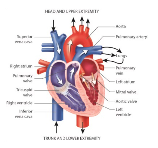
The heart is indeed made up of four chambers. The four chambers of the heart are:
- Right Atrium: This chamber receives deoxygenated blood from the body through the superior and inferior vena cava.
- Right Ventricle: The right ventricle receives the deoxygenated blood from the right atrium and pumps it to the lungs for oxygenation through the pulmonary artery.
- Left Atrium: The left atrium receives oxygenated blood from the lungs through the pulmonary veins.
- Left Ventricle: The left ventricle receives the oxygenated blood from the left atrium and pumps it out to the rest of the body through the aorta, the largest artery.
The atria and ventricles are separated by valves that ensure the flow of blood in one direction. The tricuspid valve separates the right atrium from the right ventricle, and the mitral (or bicuspid) valve separates the left atrium from the left ventricle.
These four chambers work together to ensure the continuous circulation of blood throughout the body, supplying oxygen and nutrients to the tissues and organs and removing waste products.
Heart Wall
The heart wall is composed of three layers, which are:
- Epicardium: The epicardium is the outermost layer of the heart wall. It is a thin, protective layer that also contains blood vessels, nerves, and connective tissues.
- Myocardium: The myocardium is the middle and thickest layer of the heart wall. It is made up of cardiac muscle tissue, which is responsible for the contraction and pumping action of the heart. The myocardium is well-supplied with blood vessels to provide oxygen and nutrients to the cardiac muscle cells.
- Endocardium: The endocardium is the innermost layer of the heart wall. It is a smooth, thin layer that lines the chambers of the heart and covers the heart valves. The endocardium helps to reduce friction as blood flows through the heart.
These three layers work together to maintain the structure, function, and integrity of the heart. The myocardium, with its strong contraction, allows the heart to pump blood effectively, while the epicardium and endocardium provide protection and a smooth surface for efficient blood flow.
Valves
The heart consists of four valves, which are essential for maintaining the unidirectional flow of blood through the heart. These valves are:
- Tricuspid valve: The tricuspid valve is located between the right atrium and the right ventricle. It has three cusps or flaps that open to allow blood to flow from the right atrium to the right ventricle and close to prevent backflow.
- Pulmonary valve: The pulmonary valve is situated between the right ventricle and the pulmonary artery. It consists of three semilunar cusps that open when the right ventricle contracts, allowing blood to be pumped into the pulmonary artery, and close to prevent blood from flowing back into the ventricle.
- Mitral valve: The mitral valve, also known as the bicuspid valve, is located between the left atrium and the left ventricle. It consists of two cusps and permits blood to flow from the left atrium to the left ventricle while preventing backflow.
- Aortic valve: The aortic valve is positioned between the left ventricle and the aorta. It comprises three semilunar cusps that open when the left ventricle contracts, enabling blood to be ejected into the aorta, and close to prevent blood from flowing back into the ventricle.
These valves play a crucial role in ensuring that blood flows in the correct direction through the heart chambers, allowing for efficient circulation throughout the body.
Arteries
Arteries are blood vessels that carry oxygenated blood away from the heart to various parts of the body. They form a vital part of the circulatory system and play a crucial role in delivering oxygen and nutrients to tissues and organs throughout the body. Here are some key points about arteries:
- Structure: Arteries have a thick and elastic muscular wall, allowing them to withstand the high pressure of blood being pumped from the heart. The walls are composed of three layers: the inner endothelium, the middle smooth muscle layer, and the outer connective tissue layer.
- Function: Arteries carry oxygen-rich blood from the heart to the tissues and organs of the body. They supply essential nutrients, hormones, and oxygen to these tissues, ensuring their proper functioning.
- Oxygenated Blood: Arteries carry oxygenated blood, except for the pulmonary artery, which carries deoxygenated blood from the heart to the lungs for oxygenation.
- Branching Network: Arteries branch out into smaller vessels called arterioles, which further divide into even smaller capillaries. This branching network allows for the distribution of blood to different regions and tissues.
- Blood Pressure: Arteries experience high blood pressure due to the forceful contraction of the heart, which helps propel blood throughout the body. The elasticity of the arterial walls allows them to expand and contract, maintaining a steady flow of blood.
Examples: Some examples of major arteries in the human body include the aorta, which is the largest artery and carries oxygenated blood from the heart to the rest of the body, and the carotid arteries, which supply blood to the head and neck.
It’s important to note that arteries are just one component of the circulatory system, working in conjunction with veins and capillaries to ensure proper blood circulation and oxygenation of tissues.
Veins
Veins are blood vessels that carry deoxygenated blood from various parts of the body back to the heart. They are an essential part of the circulatory system and play a crucial role in returning blood to the heart for oxygenation. Here are some key points about veins:
- Structure: Veins have thinner walls compared to arteries and have less muscular and elastic tissue. They have three layers: the inner endothelium, the middle smooth muscle layer, and the outer connective tissue layer.
- Function: Veins carry deoxygenated blood from the tissues and organs back to the heart. They collect blood that has delivered nutrients and oxygen to the tissues and return it to the heart for oxygenation and redistribution.
- Deoxygenated Blood: Veins carry deoxygenated blood, except for the pulmonary veins, which carry oxygenated blood from the lungs back to the heart.
- Low Pressure: Veins experience lower blood pressure compared to arteries. They rely on the contraction of surrounding muscles and one-way valves to help propel blood toward the heart against gravity.
- Valves: Veins contain one-way valves that prevent the backward flow of blood. These valves ensure that blood flows in the right direction, from the tissues toward the heart.
- Capacitance Vessels: Veins have a higher capacity to stretch and accommodate blood volume changes. They act as storage vessels, allowing blood to pool temporarily and regulate blood distribution during changes in body position or physical activity.
Examples: Some examples of major veins in the human body include the superior and inferior vena cava, which are the largest veins and carry deoxygenated blood from the body back to the heart, and the jugular veins, which drain blood from the head and neck.
Veins work together with arteries and capillaries to ensure the proper functioning of the circulatory system, maintaining a continuous flow of blood and facilitating the exchange of oxygen, nutrients, and waste products between the body’s cells and organs.
Frequently Asked Questions on Hearts
How does the heart function?
The heart functions as a muscular pump that circulates blood throughout the body. It receives oxygen-depleted blood from the body, pumps it to the lungs for oxygenation, and then pumps oxygen-rich blood back to the body's tissues.
What are the 7 main functions of the heart?
The heart has several vital functions: Pumping Oxygenated Blood: The heart pumps oxygen-rich blood to supply tissues. Receiving Deoxygenated Blood: It receives oxygen-depleted blood from the body. Circulation: The heart ensures a continuous flow of blood through arteries, capillaries, and veins. Oxygen Exchange: Blood gets oxygenated in the lungs and releases carbon dioxide. Nutrient Transport: The heart helps transport nutrients to body cells. Waste Removal: It aids in carrying waste products, like carbon dioxide, away from cells. Blood Pressure Regulation: The heart maintains blood pressure to support circulation.
What are the names of the parts of the heart?
The heart consists of several parts, including the atria, ventricles, valves (tricuspid, mitral, pulmonary, aortic), and the septum.
What is the structure of the heart?
The heart has four chambers: two atria (upper chambers) and two ventricles (lower chambers). Valves separate the chambers and ensure blood flows in one direction.
What is the size and shape of the heart?
The heart is roughly the size of a fist, and its shape is conical. It's situated slightly tilted to the left in the chest cavity.
What is the difference between artery and vein?
Arteries carry oxygenated blood away from the heart to the body. Veins transport oxygen-depleted blood from the body back to the heart.
What are the risk factors for heart disease?
Several risk factors contribute to heart disease: High Blood Pressure High Cholesterol Smoking Obesity Diabetes Sedentary Lifestyle Poor Diet Family History Age Stress


