Table of Contents
Introduction to Spinal Cord


The spinal cord, part of the central nervous system, contains about 100 million neurons and numerous neuroglia. It is responsible for processing reflexes and integrating excitatory and inhibitory signals. The gray matter handles reflexes and integration, while the white matter contains sensory and motor tracts that serve as “highways” for transmitting information to and from the brain. The spinal cord and brain together form the CNS.
Protection of spinal cord
The spinal cord is protected by the bony vertebral column. The three meninges that surround the spinal cord are:
- Outer thick and tough dura mater
- Middle, spiderweb-like, arachnoid mater
- Inner, thin pia mater which adheres to the surface of the brain and spinal cord
Between the meninges, there are spaces containing cerebrospinal fluid, providing a cushioning effect and further protection.


External Anatomy
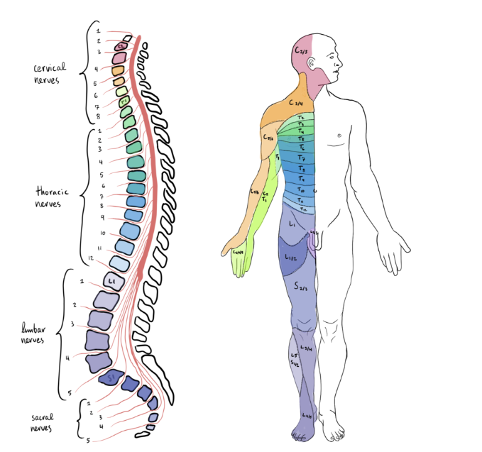
The spinal cord is oval-shaped, flattened slightly anteriorly and posteriorly, and extends from the medulla oblongata to the second lumbar vertebra in adults. It has two conspicuous enlargements – the cervical enlargement (C4 to T1) and the lumbar enlargement (from the ninth to the twelfth thoracic vertebra). Inferiorly, it tapers to form the conus medullaris, ending around the first and second lumbar vertebrae. The filum terminale, an extension of the pia mater, anchors the spinal cord to the coccyx. Spinal nerves emerge at regular intervals from the intervertebral foramina, connecting specific regions of the body to the spinal cord. The 31 pairs of spinal nerves are named based on the segments they arise from:
- 8 pairs of cervical nerves (C1–C8),
- 12 pairs of thoracic nerves (T1–T12),
- 5 pairs of lumbar nerves (L1–L5),
- 5 pairs of sacral nerves (S1–S5), and
- 1 pair of coccygeal nerves (Co1).
Each spinal nerve has a posterior (dorsal) root with sensory axons and an anterior (ventral) root with motor axons. The roots of the lower spinal nerves angle inferiorly alongside the filum terminale, collectively known as the cauda equina.
Internal anatomy
The internal anatomy of the spinal cord consists of white matter surrounding an inner core of gray matter. The white matter primarily contains bundles of myelinated axons, while the gray matter comprises dendrites, cell bodies of neurons, unmyelinated axons, and neuroglia. The gray matter is organised into functional groups called nuclei, and each side of the spinal cord is divided into regions called horns, including posterior (dorsal) gray horns, anterior (ventral) gray horns, and lateral gray horns in the thoracic and upper lumbar segments. The white matter is divided into columns, including anterior (ventral) white columns, posterior (dorsal) white columns, and lateral white columns.
The sensory input and motor output are processed within the spinal cord, involving sensory neurons conveying input from sensory receptors, and motor neurons regulating output to effector tissues.
Check for:
Spinal nerves
Spinal nerves are part of the peripheral nervous system (PNS) that connect the CNS to sensory receptors, muscles, and glands throughout the body. There are 31 pairs of spinal nerves, each named and numbered according to the region and level of the vertebral column from which they emerge.
The first cervical nerve emerges between the occipital bone and the atlas, while the remaining spinal nerves exit through the intervertebral foramina between adjacent vertebrae.
- C1-C7 (cervical nerves) exit above their corresponding vertebrae
- C8 exits between C7 and T1
- T1-L5 (Thoracic nerves) exit below their corresponding vertebrae
- S1-S5 (Sacral nerves) and Co1 (Coccygeal nerve) enter the sacral canal and exit through anterior and posterior sacral foramina or the sacral hiatus.
Each spinal nerve has a posterior root containing sensory axons and an anterior root containing motor axons, and they unite at the intervertebral foramen to form a mixed nerve. The posterior root contains a posterior root ganglion with cell bodies of sensory neurons.


Connective tissue covering
Spinal and cranial nerves are composed of individual axons wrapped in connective tissue coverings. The endoneurium surrounds individual axons, while fascicles of axons are enclosed by the perineurium. The epineurium covers the entire nerve and fuses with the dura mater of the spinal meninges at the intervertebral foramen. Spinal nerves divide into posterior and anterior rami, which serve different areas of the body. The anterior rami form plexuses, such as the cervical, brachial, lumbar, and sacral plexuses. Each spinal nerve contains sensory neurons that supply specific segments of the skin, known as dermatomes.
- Cervical plexus – It supplies the skin and muscles of the head, neck, and superior part of the shoulders and chest. It gives rise to the phrenic nerves that supply motor fibers to the diaphragm.
- Lumbar plexus – It supplies the anterolateral abdominal wall, external genitals, and part of the lower limbs.
- Sacral plexus – It supplies the buttocks, perineum, and lower limbs. The largest nerve in the body, the sciatic nerve, arises from the sacral plexus.
Coccygeal plexus is formed by the roots of spinal nerves S4–S5 and the coccygeal nerves. From this plexus, the anococcygeal nerves arise, supplying a small area of skin in the coccygeal region.
Spinal cord Functions
The spinal cord serves two main functions: nerve impulse propagation and integration of information. White matter tracts act as highways for sensory input to the brain and motor output from the brain to effector tissues. The gray matter receives and integrates incoming and outgoing information. Sensory tracts convey nerve impulses for various sensations, and motor tracts control voluntary movements. Sensory systems inform the CNS about the environment, and motor output to skeletal muscles is regulated by direct and indirect pathways. The spinal cord’s role in integrating sensory and motor information helps maintain homeostasis and coordinate responses and movements.
Reflex arc
The spinal cord serves as an integrating center for reflexes, which are fast, involuntary responses to specific stimuli. Reflex arcs consist of five components: sensory receptor, sensory neuron, integrating center, motor neuron, and effector. The stretch reflex is a monosynaptic reflex that causes muscle contraction in response to muscle stretching, protecting the muscles from overstretching. The tendon reflex is a feedback mechanism that causes muscle relaxation to prevent tendon damage due to excessive tension. The flexor reflex and crossed extensor reflex are polysynaptic reflexes involved in withdrawing a limb from a painful stimulus and maintaining balance, respectively.
Summary of Spinal Cord
The spinal cord, part of the central nervous system, contains neurons and neuroglia responsible for reflex processing and signal integration. It is protected by the bony vertebral column and surrounded by the dura, arachnoid, and pia mater. The spinal cord’s external anatomy includes cervical and lumbar enlargements, and it tapers to the conus medullaris, anchored by the filum terminale. Spinal nerves emerge from the cord and form mixed nerves with both sensory and motor components. Connective tissue coverings, including endoneurium, perineurium, and epineurium, protect the nerves. Spinal nerves give rise to plexuses, which supply specific regions of the body. The spinal cord functions in nerve impulse propagation and integration, with sensory and motor tracts as pathways for information to and from the brain. Reflexes, such as stretch, tendon, flexor, and crossed extensor reflexes, involve sensory receptors, neurons, and effectors in quick, involuntary responses.
Frequently Asked Questions on Spinal Cord
What is the spinal cord, and what is its function?
The spinal cord is a vital part of the central nervous system (CNS). It is a bundle of nerves that extends from the brainstem down through the vertebral column. Its main functions include transmitting nerve impulses between the brain and the rest of the body and integrating sensory and motor information.
How is the spinal cord protected?
The spinal cord is protected by the bony vertebral column, which surrounds it and provides a sturdy physical barrier. Additionally, the spinal cord is covered by three layers of meninges: the dura mater, arachnoid mater, and pia mater. These layers help cushion and protect the delicate neural tissues.
What are spinal nerves, and how do they connect to the spinal cord?
Spinal nerves are part of the peripheral nervous system (PNS) and are responsible for connecting the CNS to sensory receptors, muscles, and glands throughout the body. There are 31 pairs of spinal nerves that emerge from the spinal cord through openings called intervertebral foramina. Each spinal nerve has both a posterior (dorsal) root, which contains sensory axons, and an anterior (ventral) root, which contains motor axons. These roots join together to form a mixed nerve.
What are plexuses, and what is their role in the body?
Plexuses are networks formed by the anterior rami of spinal nerves. They serve as important distribution centers, supplying specific regions of the body with sensory and motor innervation. The major plexuses include the cervical, brachial, lumbar, and sacral plexuses, each serving distinct areas of the body.
What is the internal anatomy of the spinal cord?
The internal anatomy of the spinal cord consists of white matter surrounding an inner core of gray matter. The white matter contains bundles of myelinated axons that form tracts, while the gray matter contains cell bodies of neurons, dendrites, unmyelinated axons, and neuroglia. The gray matter is organized into functional groups called nuclei, and each side of the spinal cord is divided into regions called horns.
What are reflex arcs, and how do they work?
Reflex arcs are neural pathways that mediate reflex actions in response to specific stimuli. They consist of five components: sensory receptors, sensory neurons, integrating centers (usually in the spinal cord), motor neurons, and effectors. When a sensory receptor is stimulated, it sends a signal through the sensory neuron to the integrating center. The integrating center processes the information and immediately sends a signal through the motor neuron to the effector, which produces the reflexive response.
What is the role of the spinal cord in sensory and motor processing?
The spinal cord plays a crucial role in sensory and motor processing. Sensory tracts in the spinal cord convey nerve impulses for various sensations, such as pain, touch, and temperature, from the body to the brain. Motor tracts, on the other hand, carry nerve impulses from the brain to muscles, controlling voluntary movements. The spinal cord also contains interneurons that integrate sensory input and coordinate motor output, contributing to reflex actions and rapid responses to stimuli.
How does the spinal cord contribute to maintaining homeostasis?
The spinal cord, as part of the CNS, helps maintain homeostasis by processing sensory information from the body and coordinating motor responses to maintain a stable internal environment. For example, reflexes such as the stretch reflex and tendon reflex help regulate muscle tension and prevent overstretching or damage to tendons. The spinal cord's ability to integrate information allows for coordinated responses to environmental changes, ensuring the body's proper functioning.